5E1J image
Deposition Date 2015-09-29
Release Date 2015-12-16
Last Version Date 2024-03-06
Entry Detail
PDB ID:
5E1J
Keywords:
Title:
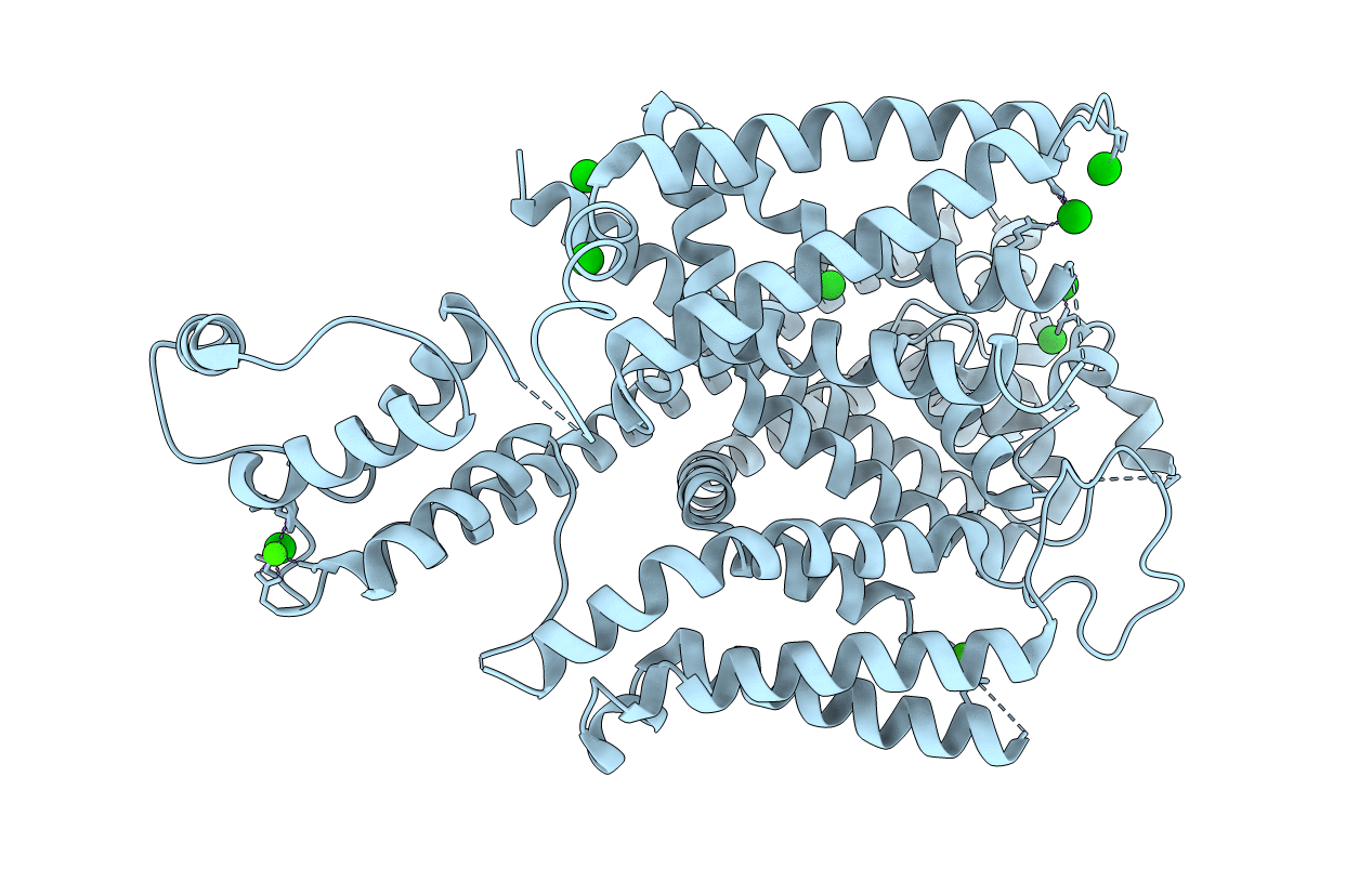
Structure of voltage-gated two-pore channel TPC1 from Arabidopsis thaliana
Biological Source:
Source Organism:
Host Organism:
Method Details:
Experimental Method:
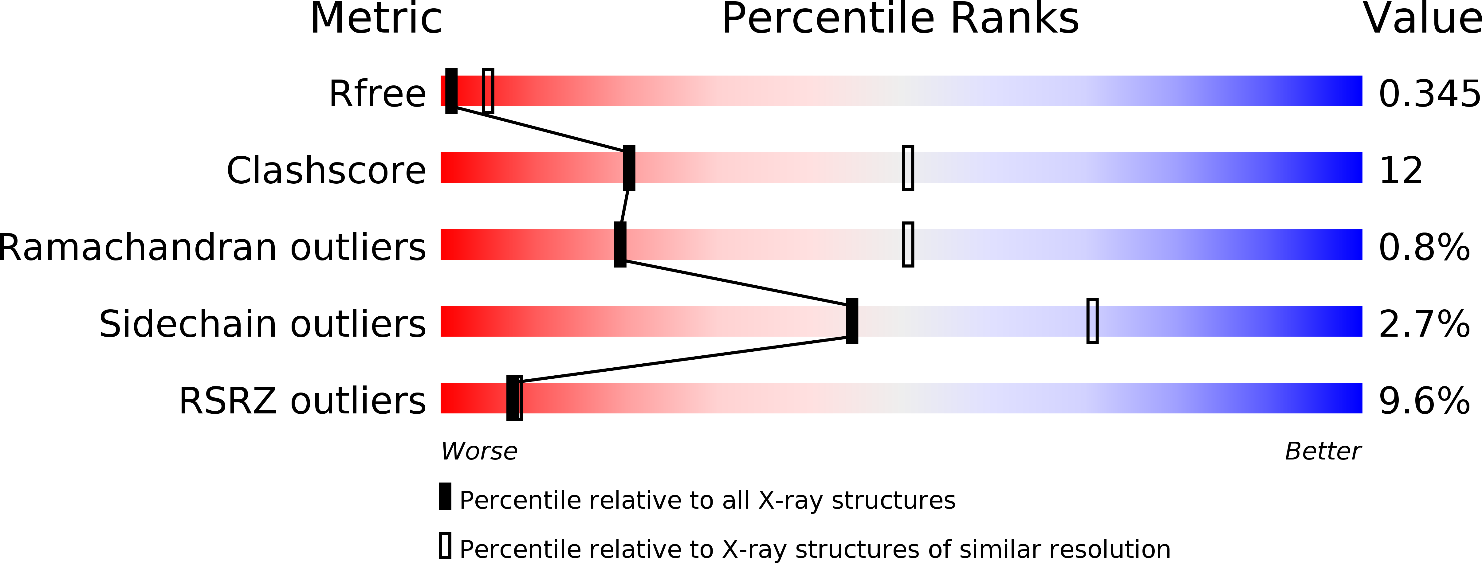
Resolution:
3.31 Å
R-Value Free:
0.33
R-Value Work:
0.32
R-Value Observed:
0.32
Space Group:
C 2 2 21
Macromolecular Entities
Structures with similar UniProt ID
Protein Blast
Polymer Type:polypeptide(L)
Molecule:Two pore calcium channel protein 1
Gene (Uniprot):TPC1
Chain IDs:A
Chain Length:741
Number of Molecules:1
Biological Source:Arabidopsis thaliana
Primary Citation
Structure of the voltage-gated two-pore channel TPC1 from Arabidopsis thaliana.
Nature 531 196 201 (2016)
PMID: 26689363 DOI: 10.1038/nature16446

Abstact

Two-pore channels (TPCs) contain two copies of a Shaker-like six-transmembrane (6-TM) domain in each subunit and are ubiquitously expressed in both animals and plants as organellar cation channels. Here we present the crystal structure of a vacuolar two-pore channel from Arabidopsis thaliana, AtTPC1, which functions as a homodimer. AtTPC1 activation requires both voltage and cytosolic Ca(2+). Ca(2+) binding to the cytosolic EF-hand domain triggers conformational changes coupled to the pair of pore-lining inner helices from the first 6-TM domains, whereas membrane potential only activates the second voltage-sensing domain, the conformational changes of which are coupled to the pair of inner helices from the second 6-TM domains. Luminal Ca(2+) or Ba(2+) can modulate voltage activation by stabilizing the second voltage-sensing domain in the resting state and shift voltage activation towards more positive potentials. Our Ba(2+)-bound AtTPC1 structure reveals a voltage sensor in the resting state, providing hitherto unseen structural insight into the general voltage-gating mechanism among voltage-gated channels.

Legend

Protein

Chemical

Disease

Primary Citation of related structures