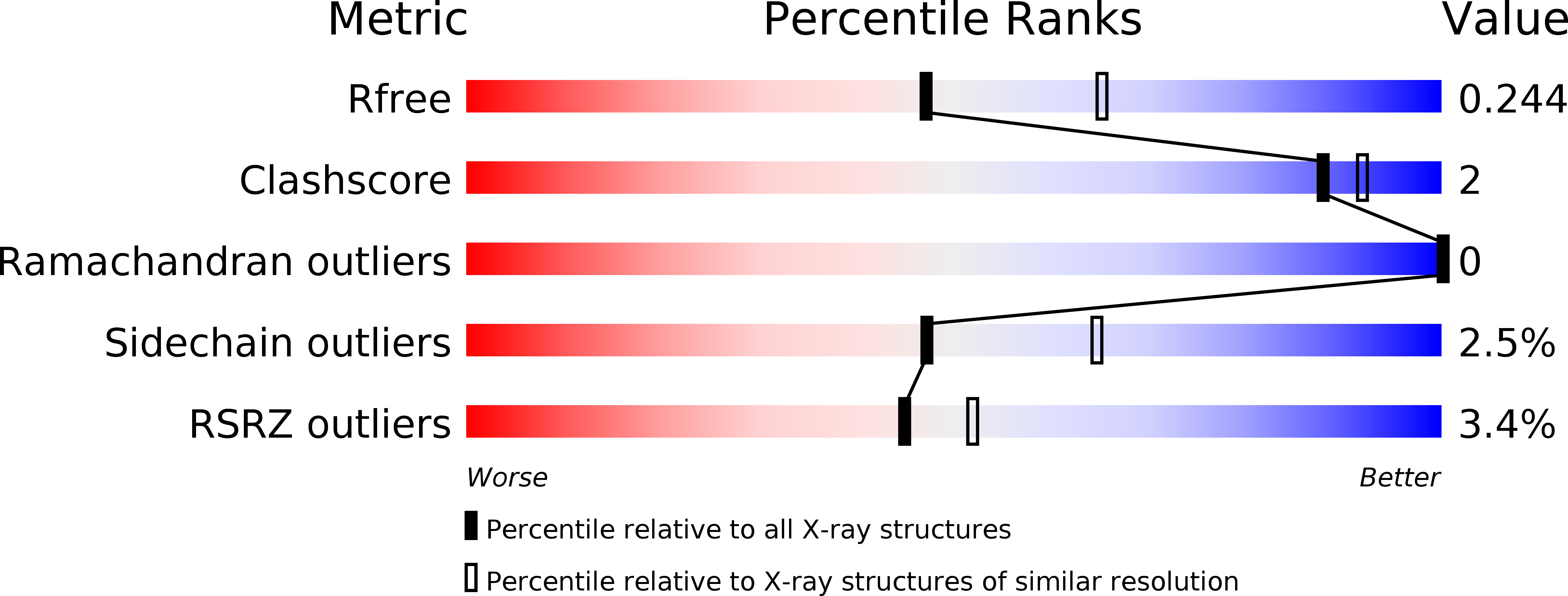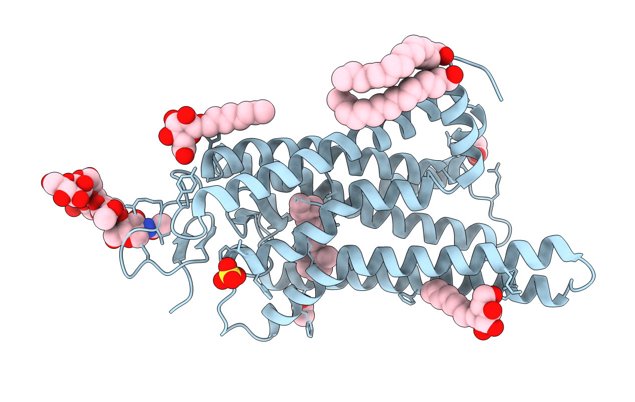
5DYS image
Entry Detail
PDB ID:
5DYS
Title:
Crystal Structure of T94I rhodopsin mutant
Biological Source:
Source Organism:
Host Organism:
PDB Version:
Deposition Date:
2015-09-25
Release Date:
2016-08-10
Method Details:
Experimental Method:
Resolution:
2.30 Å
R-Value Free:
0.23
R-Value Work:
0.21
R-Value Observed:
0.21
Space Group:
H 3 2
Macromolecular Entities
Polymer Type:polypeptide(L)
Description:Rhodopsin
Mutations:N2C, D282C, T94I
Chain IDs:A
Chain Length:349
Number of Molecules:1
Biological Source:Bos taurus
Primary Citation
Structural role of the T94I rhodopsin mutation in congenital stationary night blindness.
Embo Rep. 17 1431 1440 (2016)
PMID: 27458239 DOI: 10.15252/embr.201642671

Abstact

Congenital stationary night blindness (CSNB) is an inherited and non-progressive retinal dysfunction. Here, we present the crystal structure of CSNB-causing T94I2.61 rhodopsin in the active conformation at 2.3 Å resolution. The introduced hydrophobic side chain prolongs the lifetime of the G protein activating metarhodopsin-II state by establishing a direct van der Waals contact with K2967.43, the site of retinal attachment. This is in stark contrast to the light-activated state of the CSNB-causing G90D2.57 mutation, where the charged mutation forms a salt bridge with K2967.43 To find the common denominator between these two functional modifications, we combined our structural data with a kinetic biochemical analysis and molecular dynamics simulations. Our results indicate that both the charged G90D2.57 and the hydrophobic T94I2.61 mutation alter the dark state by weakening the interaction between the Schiff base (SB) and its counterion E1133.28 We propose that this interference with the tight regulation of the dim light photoreceptor rhodopsin increases background noise in the visual system and causes the loss of night vision characteristic for CSNB patients.

Legend

Protein

Chemical

Disease

Primary Citation of related structures