Deposition Date
2015-09-24
Release Date
2016-01-13
Last Version Date
2024-01-10
Entry Detail
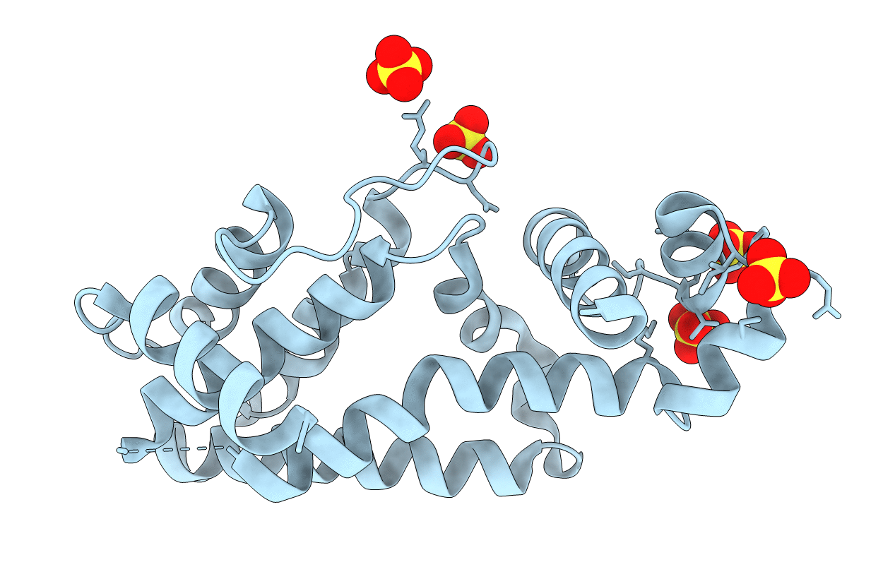
Biological Source:
Source Organism(s):
Corynebacterium glutamicum ATCC 14067 (Taxon ID: 1079988)
Expression System(s):
Method Details:
Experimental Method:
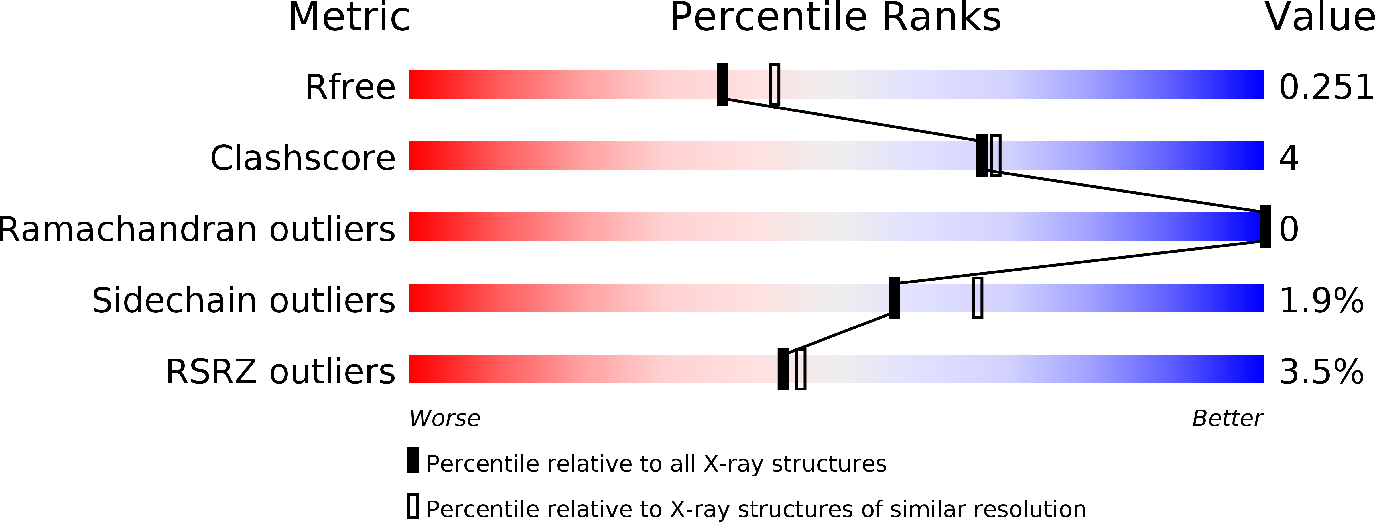
Resolution:
2.25 Å
R-Value Free:
0.24
R-Value Work:
0.21
R-Value Observed:
0.22
Space Group:
P 32 2 1