5DVI image
Deposition Date 2015-09-21
Release Date 2016-02-24
Last Version Date 2024-11-13
Entry Detail
PDB ID:
5DVI
Title:
High resolution crystal Structure of glucose complexed periplasmic glucose binding protein (ppGBP) from P. putida CSV86
Biological Source:
Source Organism:
Method Details:
Experimental Method:
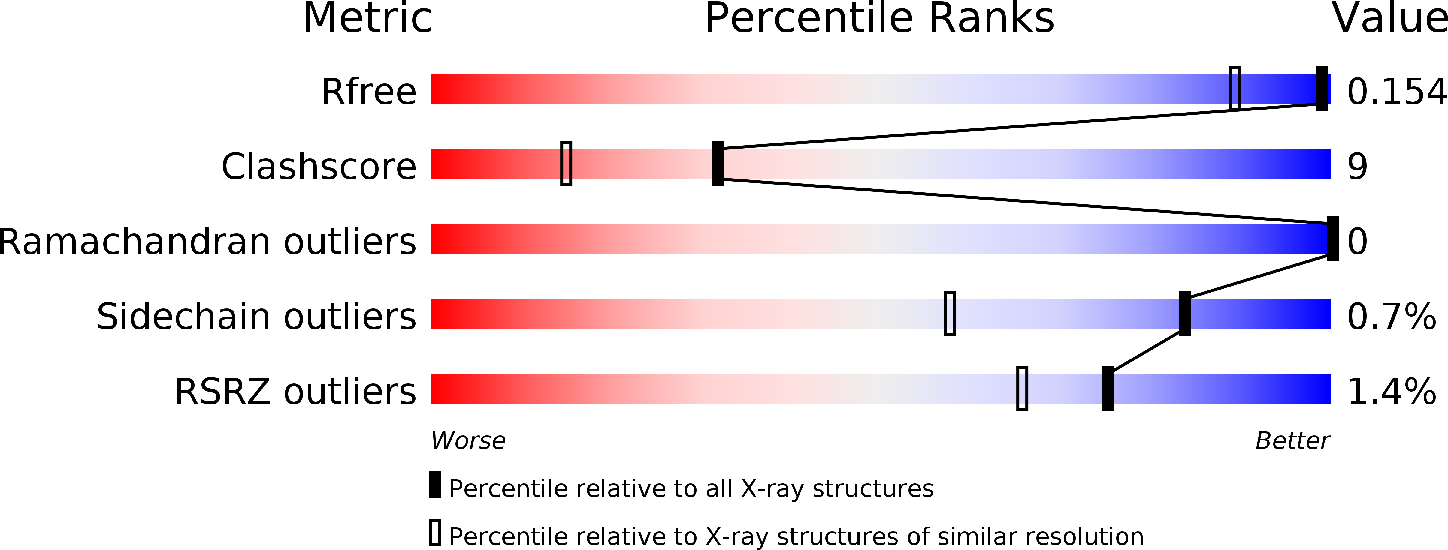
Resolution:
1.25 Å
R-Value Free:
0.15
R-Value Work:
0.12
R-Value Observed:
0.12
Space Group:
P 21 21 2
Macromolecular Entities
Structures with similar UniProt ID
Protein Blast
Polymer Type:polypeptide(L)
Molecule:Binding protein component of ABC sugar transporter
Gene (Uniprot):CSV86_019950
Chain IDs:A, B
Chain Length:419
Number of Molecules:2
Biological Source:Pseudomonas putida CSV86
Primary Citation
High Resolution Structures of Periplasmic Glucose-binding Protein of Pseudomonas putida CSV86 Reveal Structural Basis of Its Substrate Specificity
J.Biol.Chem. 291 7844 7857 (2016)
PMID: 26861882 DOI: 10.1074/jbc.M115.697268

Abstact

Periplasmic substrate-binding proteins (SBPs) bind to the specific ligand with high affinity and mediate their transport into the cytoplasm via the cognate inner membrane ATP-binding cassette proteins. Because of low sequence identities, understanding the structural basis of substrate recognition by SBPs has remained very challenging. There are several structures available for the ligand-bound sugar SBPs, but very few unliganded structures are reported. No structural data are available for sugar SBPs fromPseudomonassp. to date. This study reports the first high resolution crystal structures of periplasmic glucose-binding protein fromPseudomonas putidaCSV86 (ppGBP) in unliganded form (2.5 Å) and complexed with glucose (1.25 Å) and galactose (1.8 Å). Asymmetric domain closure of ppGBP was observed upon substrate binding. The ppGBP was found to have an affinity of ∼ 0.3 μmfor glucose. The structural analysis showed that the sugars are bound to the protein mainly by hydrogen bonds, and the loss of two strong hydrogen bonds between ppGBP and galactose compared with glucose may be responsible for lowering its affinity toward galactose. The higher stability of ppGBP-glucose complex was also indicated by an 8 °C increase in the melting temperature compared with unliganded form and ppGBP-galactose complex. ppGBP binds to monosaccharide, but the structural features revealed it to have an oligosaccharide-binding protein fold, indicating that during evolution the sugar binding pocket may have undergone structural modulation to accommodate monosaccharide only.

Legend

Protein

Chemical

Disease

Primary Citation of related structures