Deposition Date
2015-09-10
Release Date
2016-01-20
Last Version Date
2024-03-06
Entry Detail
PDB ID:
5DO9
Keywords:
Title:
Structure of regulator of G protein signaling 8 (RGS8) in complex with AlF4-activated Galpha-q
Biological Source:
Source Organism(s):
Mus musculus (Taxon ID: 10090)
Homo sapiens (Taxon ID: 9606)
Homo sapiens (Taxon ID: 9606)
Expression System(s):
Method Details:
Experimental Method:
Resolution:
2.60 Å
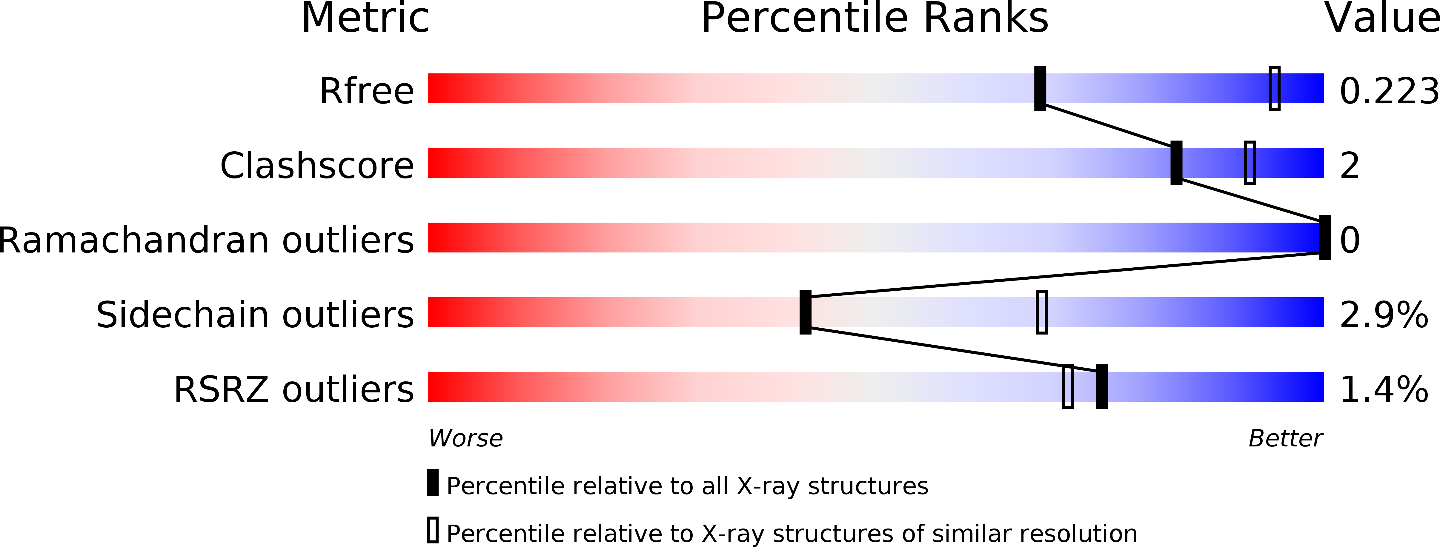
R-Value Free:
0.22
R-Value Work:
0.17
R-Value Observed:
0.18
Space Group:
C 1 2 1