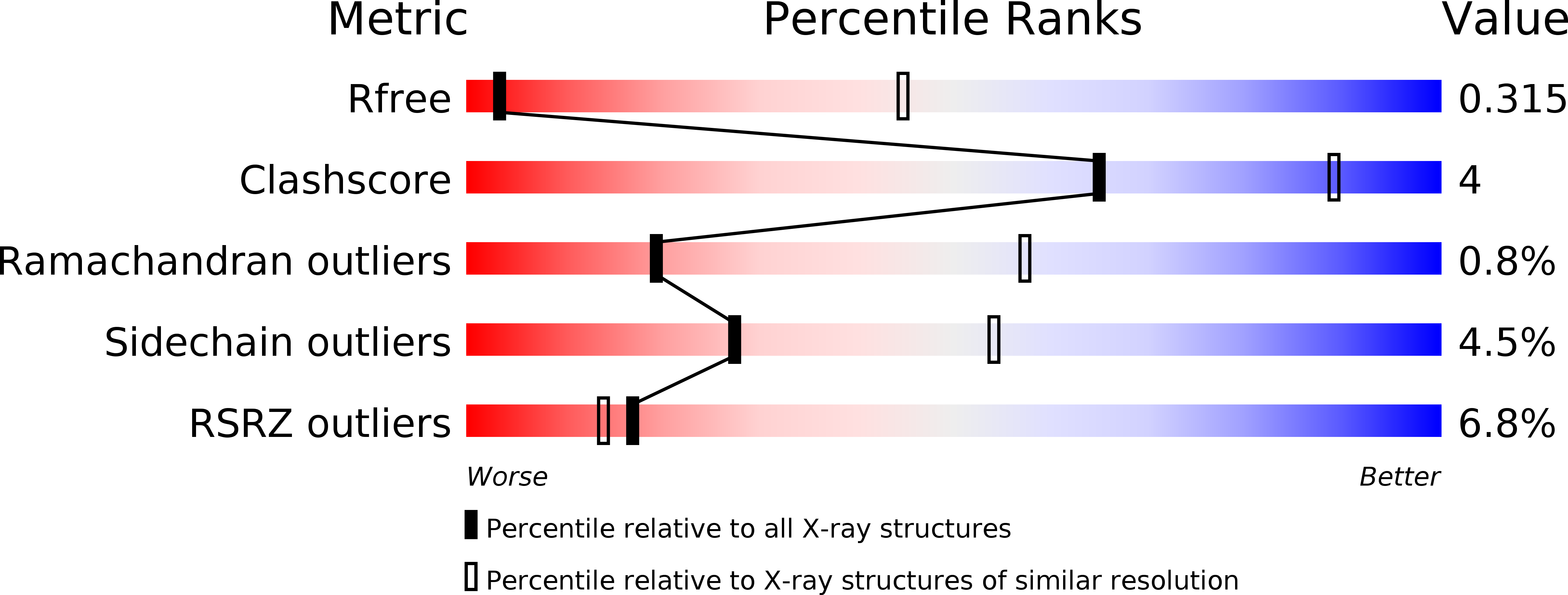
5DN6 image
Deposition Date 2015-09-09
Release Date 2015-10-28
Last Version Date 2024-05-08
Entry Detail
PDB ID:
5DN6
Keywords:
Title:
ATP synthase from Paracoccus denitrificans
Biological Source:
Source Organism:
Method Details:
Experimental Method:
Resolution:
3.98 Å
R-Value Free:
0.32
R-Value Work:
0.29
R-Value Observed:
0.29
Space Group:
P 1 21 1
Macromolecular Entities
Polymer Type:polypeptide(L)
Molecule:Chain A
Chain IDs:A (auth: 1)
Chain Length:20
Number of Molecules:1
Biological Source:Paracoccus denitrificans
Polymer Type:polypeptide(L)
Molecule:Chain B
Chain IDs:B (auth: 2)
Chain Length:15
Number of Molecules:1
Biological Source:Paracoccus denitrificans
Polymer Type:polypeptide(L)
Molecule:Chain C
Chain IDs:C (auth: 3)
Chain Length:19
Number of Molecules:1
Biological Source:Paracoccus denitrificans
Structures with similar UniProt ID
Protein Blast
Polymer Type:polypeptide(L)
Molecule:ATP synthase subunit alpha
Gene (Uniprot):atpA
Chain IDs:D (auth: A), E (auth: B), F (auth: C)
Chain Length:511
Number of Molecules:3
Biological Source:Paracoccus denitrificans
Structures with similar UniProt ID
Protein Blast
Polymer Type:polypeptide(L)
Molecule:ATP synthase subunit beta
Gene (Uniprot):atpD
Chain IDs:G (auth: D), H (auth: E), I (auth: F)
Chain Length:474
Number of Molecules:3
Biological Source:Paracoccus denitrificans
Structures with similar UniProt ID
Protein Blast
Polymer Type:polypeptide(L)
Molecule:ATP synthase gamma chain
Gene (Uniprot):atpG
Chain IDs:J (auth: G)
Chain Length:290
Number of Molecules:1
Biological Source:Paracoccus denitrificans
Structures with similar UniProt ID
Protein Blast
Polymer Type:polypeptide(L)
Molecule:ATP synthase subunit delta
Gene (Uniprot):atpH
Chain IDs:K (auth: H)
Chain Length:188
Number of Molecules:1
Biological Source:Paracoccus denitrificans
Structures with similar UniProt ID
Protein Blast
Polymer Type:polypeptide(L)
Molecule:ATP synthase epsilon chain
Gene (Uniprot):atpC
Chain IDs:L (auth: I)
Chain Length:148
Number of Molecules:1
Biological Source:Paracoccus denitrificans
Structures with similar UniProt ID
Protein Blast
Polymer Type:polypeptide(L)
Molecule:ATP synthase F0 subcomplex C subunit
Gene (Uniprot):atpE
Chain IDs:M (auth: J), N (auth: K), O (auth: L), P (auth: M), Q (auth: N), R (auth: O), S (auth: P), T (auth: Q), U (auth: R), V (auth: S), W (auth: T), X (auth: U)
Chain Length:77
Number of Molecules:12
Biological Source:Paracoccus denitrificans
Polymer Type:polypeptide(L)
Molecule:Chain V
Chain IDs:Y (auth: V)
Chain Length:78
Number of Molecules:1
Biological Source:Paracoccus denitrificans
Polymer Type:polypeptide(L)
Molecule:Chain W
Chain IDs:Z (auth: W)
Chain Length:124
Number of Molecules:1
Biological Source:Paracoccus denitrificans
Structures with similar UniProt ID
Protein Blast
Polymer Type:polypeptide(L)
Molecule:ATP synthase subunit a,ATP synthase subunit a,ATP synthase subunit a,ATP synthase subunit a
Gene (Uniprot):atpB
Chain IDs:AA (auth: X)
Chain Length:283
Number of Molecules:1
Biological Source:Paracoccus denitrificans
Polymer Type:polypeptide(L)
Molecule:Chain Y
Chain IDs:BA (auth: Y)
Chain Length:54
Number of Molecules:1
Biological Source:Paracoccus denitrificans
Structures with similar UniProt ID
Protein Blast
Polymer Type:polypeptide(L)
Molecule:Zeta inhibitor protein
Gene (Uniprot):Pden_2862
Chain IDs:CA (auth: Z)
Chain Length:104
Number of Molecules:1
Biological Source:Paracoccus denitrificans
Primary Citation
Structure of ATP synthase from Paracoccus denitrificans determined by X-ray crystallography at 4.0 angstrom resolution.
Proc.Natl.Acad.Sci.USA 112 13231 13236 (2015)
PMID: 26460036 DOI: 10.1073/pnas.1517542112

Abstact

The structure of the intact ATP synthase from the α-proteobacterium Paracoccus denitrificans, inhibited by its natural regulatory ζ-protein, has been solved by X-ray crystallography at 4.0 Å resolution. The ζ-protein is bound via its N-terminal α-helix in a catalytic interface in the F1 domain. The bacterial F1 domain is attached to the membrane domain by peripheral and central stalks. The δ-subunit component of the peripheral stalk binds to the N-terminal regions of two α-subunits. The stalk extends via two parallel long α-helices, one in each of the related b and b' subunits, down a noncatalytic interface of the F1 domain and interacts in an unspecified way with the a-subunit in the membrane domain. The a-subunit lies close to a ring of 12 c-subunits attached to the central stalk in the F1 domain, and, together, the central stalk and c-ring form the enzyme's rotor. Rotation is driven by the transmembrane proton-motive force, by a mechanism where protons pass through the interface between the a-subunit and c-ring via two half-channels in the a-subunit. These half-channels are probably located in a bundle of four α-helices in the a-subunit that are tilted at ∼30° to the plane of the membrane. Conserved polar residues in the two α-helices closest to the c-ring probably line the proton inlet path to an essential carboxyl group in the c-subunit in the proton uptake site and a proton exit path from the proton release site. The structure has provided deep insights into the workings of this extraordinary molecular machine.

Legend

Protein

Chemical

Disease

Primary Citation of related structures