5DMQ image
Deposition Date 2015-09-09
Release Date 2016-07-06
Last Version Date 2024-03-20
Entry Detail
PDB ID:
5DMQ
Title:
Crystal structure of mouse eRF1 in complex with Reverse Transcriptase (RT) of Moloney Murine Leukemia Virus
Biological Source:
Host Organism:
Method Details:
Experimental Method:
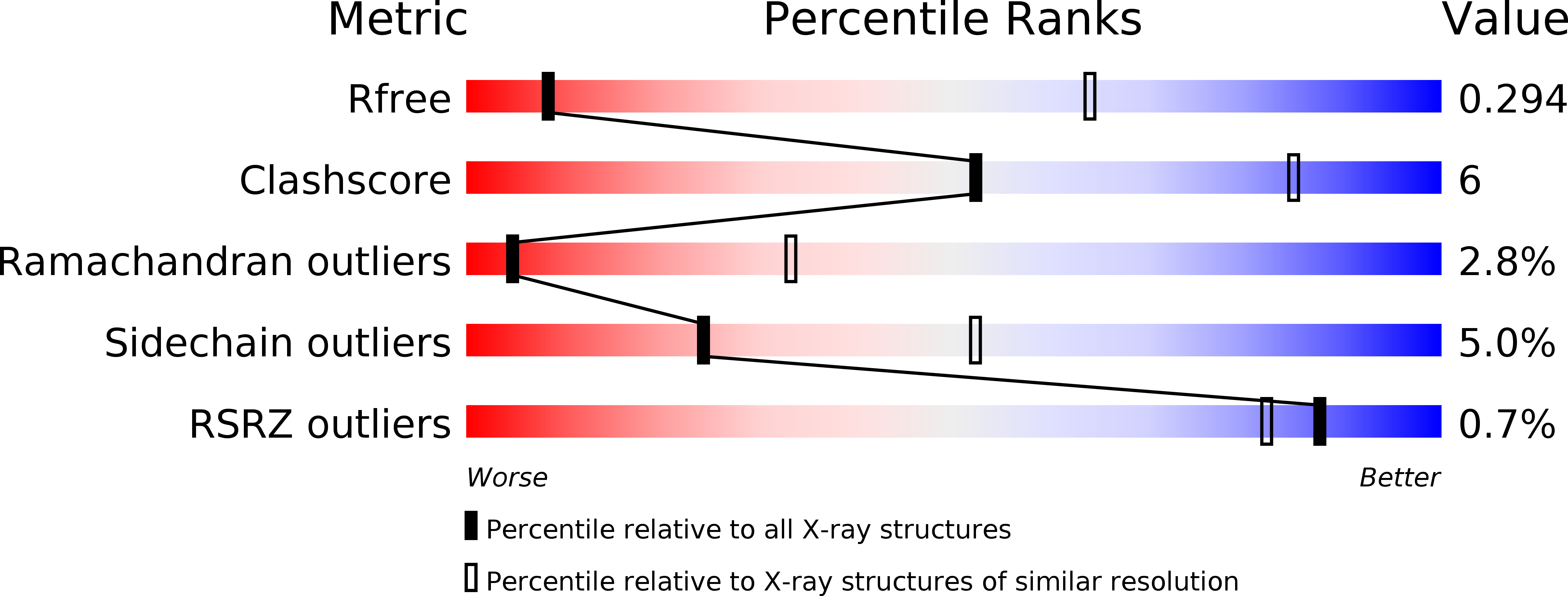
Resolution:
4.00 Å
R-Value Free:
0.29
R-Value Work:
0.24
R-Value Observed:
0.24
Space Group:
P 32 2 1
Macromolecular Entities
Polymer Type:polypeptide(L)
Molecule:Reverse transcriptase/ribonuclease H p80
Gene (Uniprot):gag-pol
Chain IDs:A
Chain Length:648
Number of Molecules:1
Biological Source:Moloney murine leukemia virus (isolate Shinnick)
Polymer Type:polypeptide(L)
Molecule:Eukaryotic peptide chain release factor subunit 1
Gene (Uniprot):Etf1
Chain IDs:B
Chain Length:437
Number of Molecules:1
Biological Source:Mus musculus
Ligand Molecules
Primary Citation
Structural basis of suppression of host translation termination by Moloney Murine Leukemia Virus
Nat Commun 7 12070 12070 (2016)
PMID: 27329342 DOI: 10.1038/ncomms12070

Abstact

Retroviral reverse transcriptase (RT) of Moloney murine leukemia virus (MoMLV) is expressed in the form of a large Gag-Pol precursor protein by suppression of translational termination in which the maximal efficiency of stop codon read-through depends on the interaction between MoMLV RT and peptidyl release factor 1 (eRF1). Here, we report the crystal structure of MoMLV RT in complex with eRF1. The MoMLV RT interacts with the C-terminal domain of eRF1 via its RNase H domain to sterically occlude the binding of peptidyl release factor 3 (eRF3) to eRF1. Promotion of read-through by MoMLV RNase H prevents nonsense-mediated mRNA decay (NMD) of mRNAs. Comparison of our structure with that of HIV RT explains why HIV RT cannot interact with eRF1. Our results provide a mechanistic view of how MoMLV manipulates the host translation termination machinery for the synthesis of its own proteins.

Legend

Protein

Chemical

Disease

Primary Citation of related structures