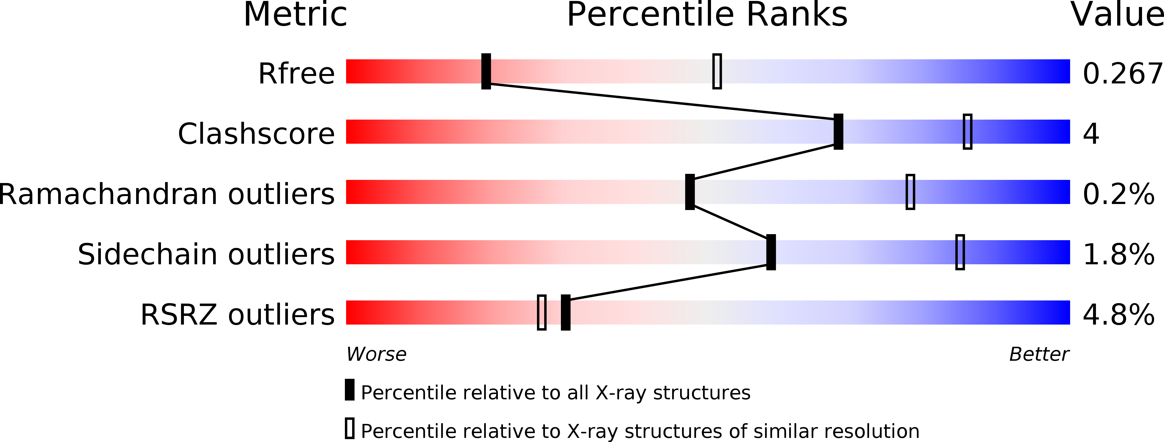
5DMN image
Deposition Date 2015-09-09
Release Date 2015-11-25
Last Version Date 2023-09-27
Entry Detail
PDB ID:
5DMN
Keywords:
Title:
Crystal Structure of the Homocysteine Methyltransferase MmuM from Escherichia coli, Apo form
Biological Source:
Source Organism:
Host Organism:
Method Details:
Experimental Method:
Resolution:
2.89 Å
R-Value Free:
0.27
R-Value Work:
0.22
R-Value Observed:
0.22
Space Group:
P 21 21 2
Macromolecular Entities
Polymer Type:polypeptide(L)
Molecule:Homocysteine S-methyltransferase
Gene (Uniprot):mmuM
Chain IDs:A, B
Chain Length:310
Number of Molecules:2
Biological Source:Escherichia coli (strain K12)
Ligand Molecules
Primary Citation
Crystal structure of the homocysteine methyltransferase MmuM from Escherichia coli.
Biochem.J. 473 277 284 (2016)
PMID: 26564203 DOI: 10.1042/BJ20150980

Abstact

Homocysteine S-methyltransferases (HMTs, EC 2.1.1.0) catalyse the conversion of homocysteine to methionine using S-methylmethionine or S-adenosylmethionine as the methyl donor. HMTs play an important role in methionine biosynthesis and are widely distributed among micro-organisms, plants and animals. Additionally, HMTs play a role in metabolite repair of S-adenosylmethionine by removing an inactive diastereomer from the pool. The mmuM gene product from Escherichia coli is an archetypal HMT family protein and contains a predicted zinc-binding motif in the enzyme active site. In the present study, we demonstrate X-ray structures for MmuM in oxidized, apo and metallated forms, representing the first such structures for any member of the HMT family. The structures reveal a metal/substrate-binding pocket distinct from those in related enzymes. The presented structure analysis and modelling of co-substrate interactions provide valuable insight into the function of MmuM in both methionine biosynthesis and cofactor repair.

Legend

Protein

Chemical

Disease

Primary Citation of related structures