
Deposition Date
2015-09-02
Release Date
2015-11-18
Last Version Date
2024-11-13
Entry Detail
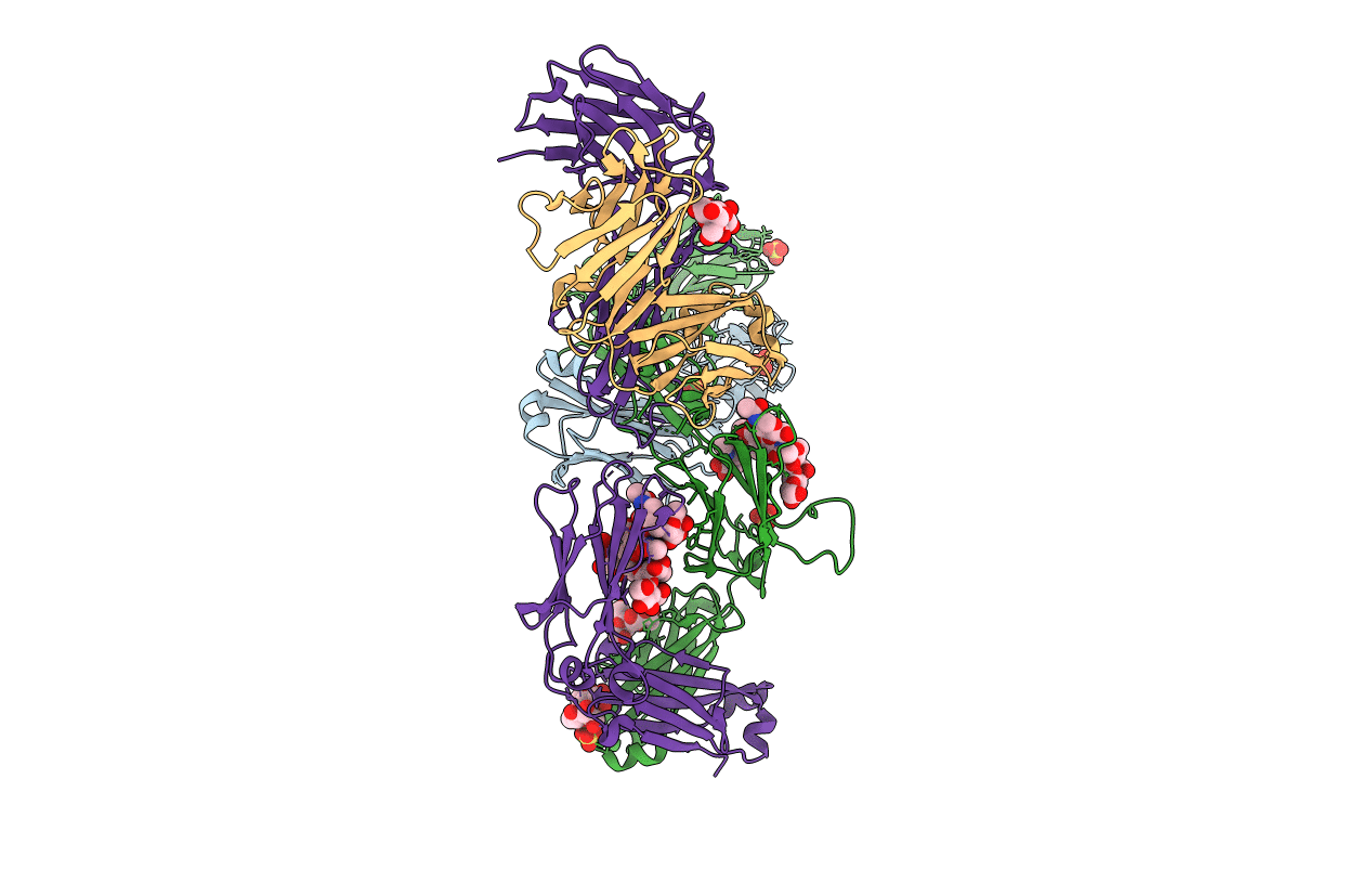
PDB ID:
5DK3
Keywords:
Title:
Crystal Structure of Pembrolizumab, a full length IgG4 antibody
Biological Source:
Source Organism(s):
Homo sapiens (Taxon ID: 9606)
Expression System(s):
Method Details:
Experimental Method:
Resolution:
2.28 Å
R-Value Free:
0.22
R-Value Work:
0.18
R-Value Observed:
0.18
Space Group:
P 21 21 21
