5DHV image
Deposition Date 2015-08-31
Release Date 2016-06-22
Last Version Date 2024-10-16
Entry Detail
PDB ID:
5DHV
Keywords:
Title:
HIV-1 Rev NTD dimers with variable crossing angles
Biological Source:
Source Organism:
Host Organism:
Method Details:
Experimental Method:
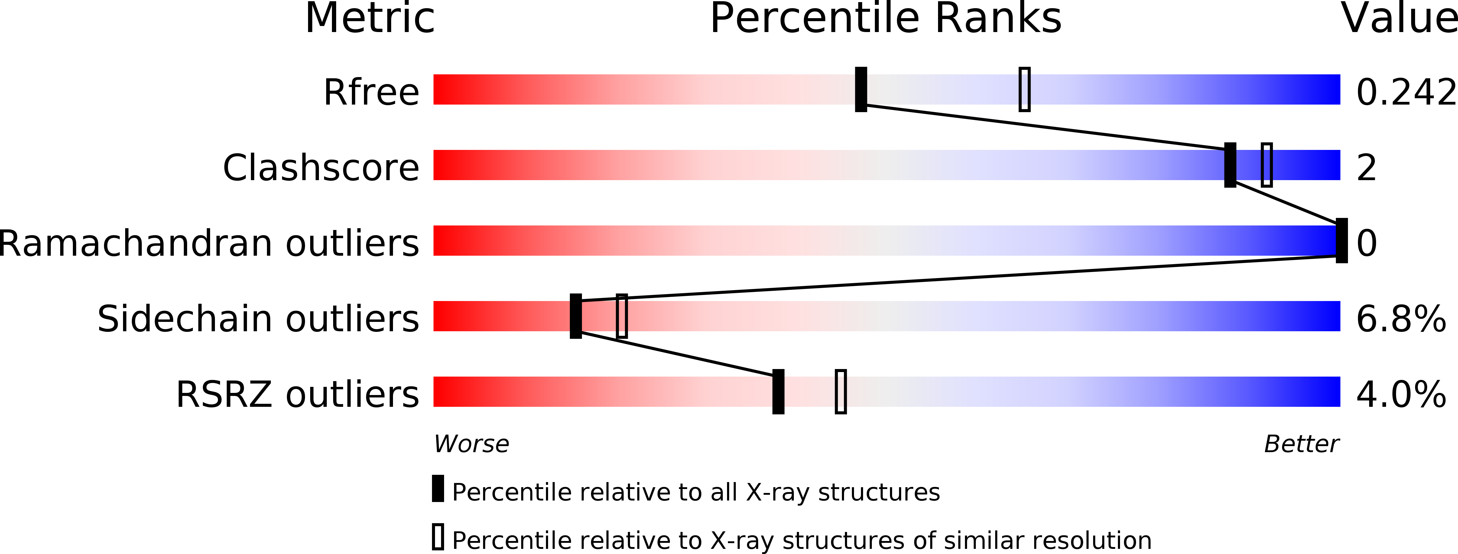
Resolution:
2.30 Å
R-Value Free:
0.22
R-Value Work:
0.18
R-Value Observed:
0.18
Space Group:
P 1 21 1
Macromolecular Entities
Polymer Type:polypeptide(L)
Molecule:Anti-Rev Antibody Fab single-chain variable fragment, heavy chain
Chain IDs:A
Chain Length:123
Number of Molecules:1
Biological Source:Oryctolagus cuniculus
Polymer Type:polypeptide(L)
Molecule:Anti-Rev Antibody Fab single-chain variable fragment, light chain
Chain IDs:B, D (auth: L)
Chain Length:110
Number of Molecules:2
Biological Source:Oryctolagus cuniculus
Polymer Type:polypeptide(L)
Molecule:Anti-Rev Antibody Fab single-chain variable fragment, heavy chain
Chain IDs:C (auth: H)
Chain Length:117
Number of Molecules:1
Biological Source:Oryctolagus cuniculus
Polymer Type:polypeptide(L)
Molecule:Protein Rev
Chain IDs:E (auth: M), F (auth: N)
Chain Length:65
Number of Molecules:2
Biological Source:Human immunodeficiency virus 1
Ligand Molecules
Primary Citation
The Structure of HIV-1 Rev Filaments Suggests a Bilateral Model for Rev-RRE Assembly.
Structure 24 1068 1080 (2016)
PMID: 27265851 DOI: 10.1016/j.str.2016.04.015

Abstact

HIV-1 Rev protein mediates the nuclear export of viral RNA genomes. To do so, Rev oligomerizes cooperatively onto an RNA motif, the Rev response element (RRE), forming a complex that engages with the host nuclear export machinery. To better understand Rev oligomerization, we determined four crystal structures of Rev N-terminal domain dimers, which show that they can pivot about their dyad axis, giving crossing angles of 90° to 140°. In parallel, we performed cryoelectron microscopy of helical Rev filaments. Filaments vary from 11 to 15 nm in width, reflecting variations in dimer crossing angle. These structures contain additional density, indicating that C-terminal domains become partially ordered in the context of filaments. This conformational variability may be exploited in the assembly of RRE/Rev complexes. Our data also revealed a third interface between Revs, which offers an explanation for how the arrangement of Rev subunits adapts to the "A"-shaped architecture of the RRE in export-active complexes.

Legend

Protein

Chemical

Disease

Primary Citation of related structures