5DG4 image
Deposition Date 2015-08-27
Release Date 2016-09-21
Last Version Date 2024-03-06
Entry Detail
PDB ID:
5DG4
Title:
Crystal structure of monomer human cellular retinol binding protein II-Y60L
Biological Source:
Source Organism:
Homo sapiens (Taxon ID: 9606)
Host Organism:
Method Details:
Experimental Method:
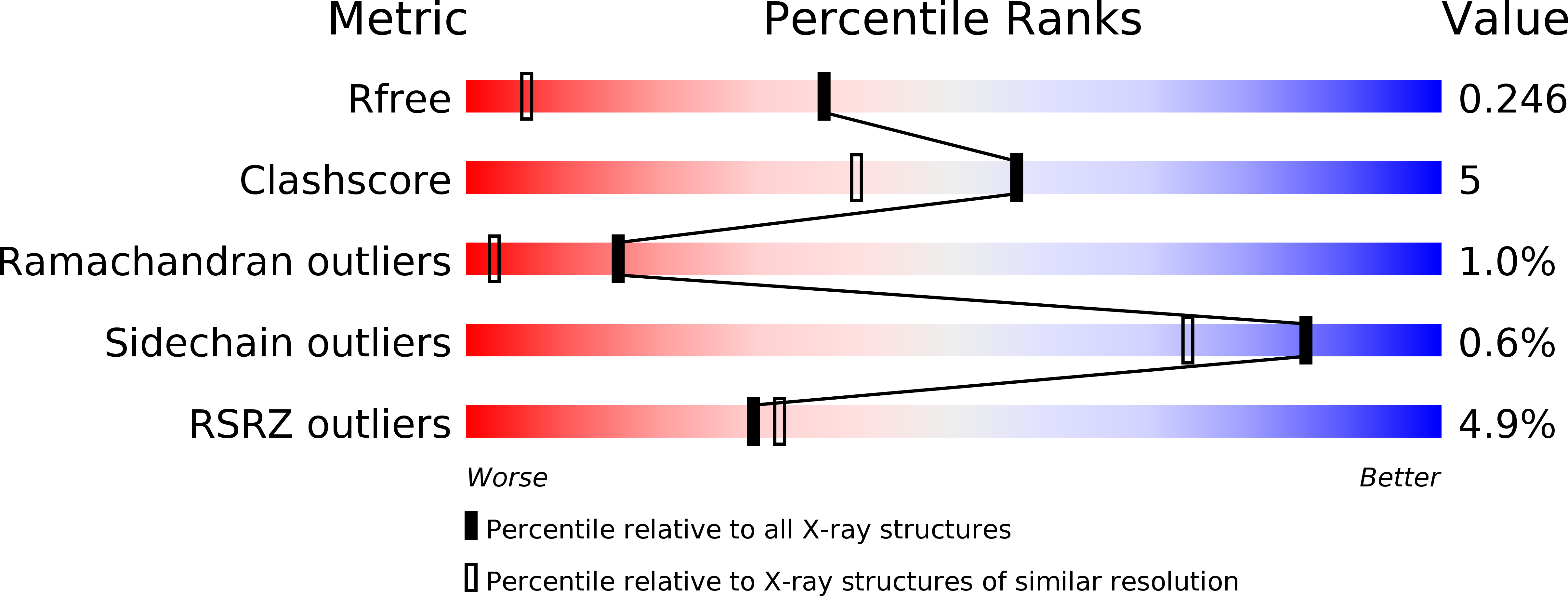
Resolution:
1.50 Å
R-Value Free:
0.24
R-Value Work:
0.22
R-Value Observed:
0.22
Space Group:
P 1
Macromolecular Entities
Polymer Type:polypeptide(L)
Molecule:Retinol-binding protein 2
Gene (Uniprot):RBP2
Mutations:Y60L
Chain IDs:A, B, C, D
Chain Length:133
Number of Molecules:4
Biological Source:Homo sapiens
Ligand Molecules
Primary Citation
Domain-Swapped Dimers of Intracellular Lipid-Binding Proteins: Evidence for Ordered Folding Intermediates.
Structure 24 1590 1598 (2016)
PMID: 27524203 DOI: 10.1016/j.str.2016.05.022

Abstact

Human Cellular Retinol Binding Protein II (hCRBPII), a member of the intracellular lipid-binding protein family, is a monomeric protein responsible for the intracellular transport of retinol and retinal. Herein we report that hCRBPII forms an extensive domain-swapped dimer during bacterial expression. The domain-swapped region encompasses almost half of the protein. The dimer represents a novel structural architecture with the mouths of the two binding cavities facing each other, producing a new binding cavity that spans the length of the protein complex. Although wild-type hCRBPII forms the dimer, the propensity for dimerization can be substantially increased via mutation at Tyr60. The monomeric form of the wild-type protein represents the thermodynamically more stable species, making the domain-swapped dimer a kinetically trapped entity. Hypothetically, the wild-type protein has evolved to minimize dimerization of the folding intermediate through a critical hydrogen bond (Tyr60-Glu72) that disfavors the dimeric form.

Legend

Protein

Chemical

Disease

Primary Citation of related structures