5DG2 image
Deposition Date 2015-08-27
Release Date 2016-09-14
Last Version Date 2023-11-08
Entry Detail
PDB ID:
5DG2
Title:
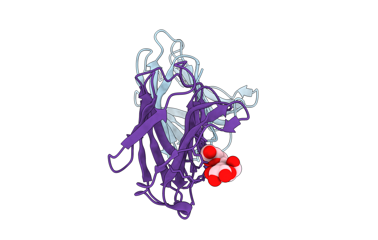
Sugar binding protein - human galectin-2 (dimer)
Biological Source:
Source Organism:
Homo sapiens (Taxon ID: 9606)
Host Organism:
Method Details:
Experimental Method:
Resolution:
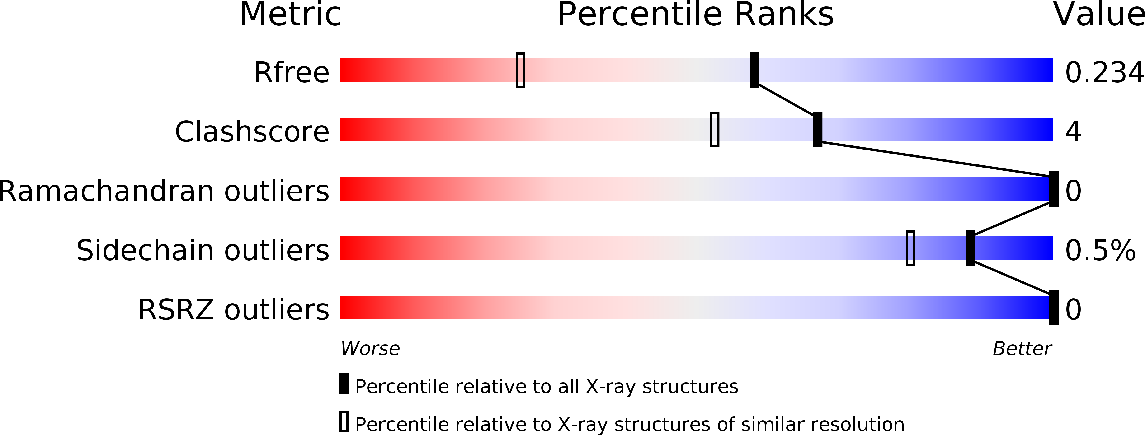
1.61 Å
R-Value Free:
0.23
R-Value Work:
0.20
R-Value Observed:
0.20
Space Group:
P 2 21 21
Macromolecular Entities
Structures with similar UniProt ID
Protein Blast
Polymer Type:polypeptide(L)
Molecule:Galectin-2
Gene (Uniprot):LGALS2
Chain IDs:A, B
Chain Length:135
Number of Molecules:2
Biological Source:Homo sapiens
Peptide-like Molecules
PRD_900008
Primary Citation
Human galectin-2 interacts with carbohydrates and peptides non-classically: new insight from X-ray crystallography and hemagglutination.
Acta Biochim. Biophys. Sin. (Shanghai) 48 939 947 (2016)
PMID: 27563008 DOI: 10.1093/abbs/gmw089

Abstact

Galectin-2 (Gal-2) plays a role in cancer, myocardial infarction, immune response, and gastrointestinal tract diseases. The only reported crystal structure of Gal-2 shows that it is a dimer in which the monomer subunits have almost identical structures, each binding with one molecule of lactose. In this study, we crystallized Gal-2 under new conditions that produced three crystal structures. In each Gal-2 dimer structure, lactose was shown to be bound to only one of the carbohydrate recognition domain subunits. In solution studies, the thermal shift assay demonstrated that inequivalent monomer subunits in the Gal-2 dimer become equivalent upon ligand binding. In addition, galectin-mediated erythrocyte agglutination assays using lactose and larger complex polysaccharides as inhibitors showed the structural differences between Gal-1 and Gal-2. Overall, our results reveal some novel aspects to the structural differentiation in Gal-2 and expand the potential for different types of molecular interactions that may be specific to this lectin.

Legend

Protein

Chemical

Disease

Primary Citation of related structures