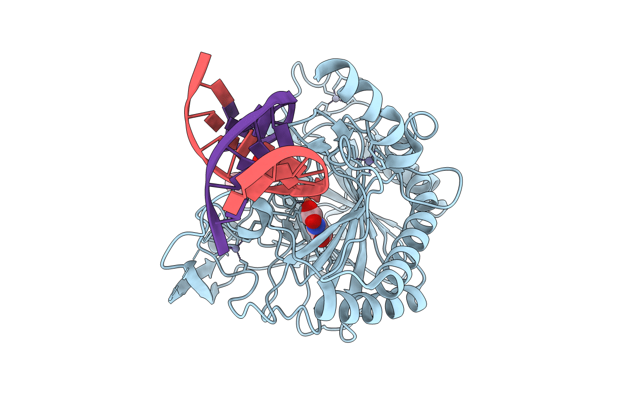
5DEU image
Entry Detail
PDB ID:
5DEU
Title:
Crystal structure of TET2-5hmC complex
Biological Source:
Host Organism:
PDB Version:
Deposition Date:
2015-08-26
Release Date:
2015-11-04
Method Details:
Experimental Method:
Resolution:
1.80 Å
R-Value Free:
0.21
R-Value Work:
0.17
R-Value Observed:
0.17
Space Group:
C 2 2 21
Macromolecular Entities
Polymer Type:polypeptide(L)
Description:Methylcytosine dioxygenase TET2, chimeric construct
Chain IDs:A
Chain Length:462
Number of Molecules:1
Biological Source:Homo sapiens
Polymer Type:polydeoxyribonucleotide
Description:DNA (5'-D(*AP*CP*CP*AP*CP*(5HC)P*GP*GP*TP*GP*GP*T)-3')
Chain IDs:B
Chain Length:12
Number of Molecules:1
Biological Source:synthetic construct
Polymer Type:polydeoxyribonucleotide
Description:DNA (5'-D(P*CP*CP*AP*CP*(5HC)P*GP*GP*TP*GP*GP*T)-3')
Chain IDs:C
Chain Length:11
Number of Molecules:1
Biological Source:synthetic construct
Primary Citation
Structural insight into substrate preference for TET-mediated oxidation.
Nature 527 118 122 (2015)
PMID: 26524525 DOI: 10.1038/nature15713

Abstact

DNA methylation is an important epigenetic modification. Ten-eleven translocation (TET) proteins are involved in DNA demethylation through iteratively oxidizing 5-methylcytosine (5mC) into 5-hydroxymethylcytosine (5hmC), 5-formylcytosine (5fC) and 5-carboxylcytosine (5caC). Here we show that human TET1 and TET2 are more active on 5mC-DNA than 5hmC/5fC-DNA substrates. We determine the crystal structures of TET2-5hmC-DNA and TET2-5fC-DNA complexes at 1.80 Å and 1.97 Å resolution, respectively. The cytosine portion of 5hmC/5fC is specifically recognized by TET2 in a manner similar to that of 5mC in the TET2-5mC-DNA structure, and the pyrimidine base of 5mC/5hmC/5fC adopts an almost identical conformation within the catalytic cavity. However, the hydroxyl group of 5hmC and carbonyl group of 5fC face towards the opposite direction because the hydroxymethyl group of 5hmC and formyl group of 5fC adopt restrained conformations through forming hydrogen bonds with the 1-carboxylate of NOG and N4 exocyclic nitrogen of cytosine, respectively. Biochemical analyses indicate that the substrate preference of TET2 results from the different efficiencies of hydrogen abstraction in TET2-mediated oxidation. The restrained conformation of 5hmC and 5fC within the catalytic cavity may prevent their abstractable hydrogen(s) adopting a favourable orientation for hydrogen abstraction and thus result in low catalytic efficiency. Our studies demonstrate that the substrate preference of TET2 results from the intrinsic value of its substrates at their 5mC derivative groups and suggest that 5hmC is relatively stable and less prone to further oxidation by TET proteins. Therefore, TET proteins are evolutionarily tuned to be less reactive towards 5hmC and facilitate the generation of 5hmC as a potentially stable mark for regulatory functions.

Legend

Protein

Chemical

Disease

Primary Citation of related structures