5DE2 image
Entry Detail
PDB ID:
5DE2
Keywords:
Title:
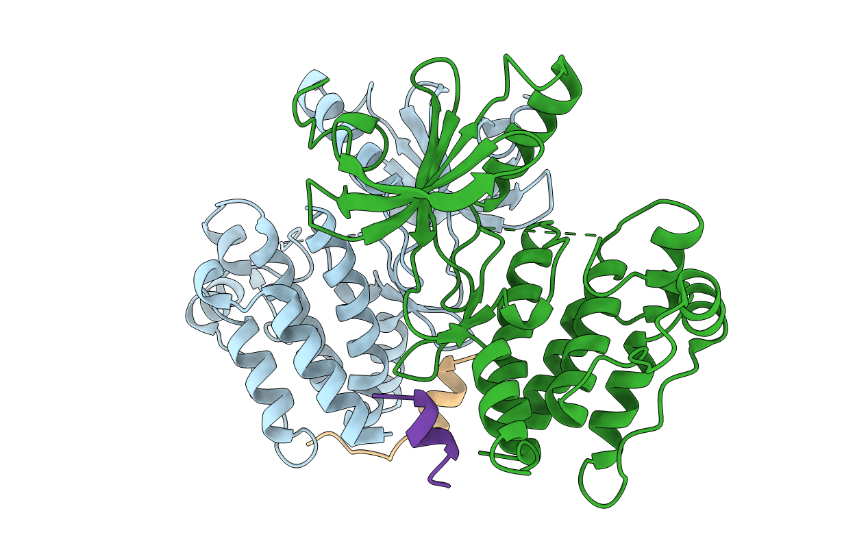
Structural mechanism of Nek7 activation by Nek9-induced dimerisation
Biological Source:
Source Organism:
Host Organism:
PDB Version:
Deposition Date:
2015-08-25
Release Date:
2015-11-11
Method Details:
Experimental Method:
Resolution:
2.78 Å
R-Value Free:
0.25
R-Value Work:
0.20
R-Value Observed:
0.20
Space Group:
P 31 2 1
Macromolecular Entities
Polymer Type:polypeptide(L)
Description:Serine/threonine-protein kinase Nek7
Mutations:Y97F
Chain IDs:A, B
Chain Length:310
Number of Molecules:2
Biological Source:Homo sapiens
Polymer Type:polypeptide(L)
Description:Serine/threonine-protein kinase Nek9
Chain IDs:C, D
Chain Length:19
Number of Molecules:2
Biological Source:Mus musculus
Primary Citation
Mechanistic basis of Nek7 activation through Nek9 binding and induced dimerization.
Nat Commun 6 8771 8771 (2015)
PMID: 26522158 DOI: 10.1038/ncomms9771

Abstact

Mitotic spindle assembly requires the regulated activities of protein kinases such as Nek7 and Nek9. Nek7 is autoinhibited by the protrusion of Tyr97 into the active site and activated by the Nek9 non-catalytic C-terminal domain (CTD). CTD binding apparently releases autoinhibition because mutation of Tyr97 to phenylalanine increases Nek7 activity independently of Nek9. Here we find that self-association of the Nek9-CTD is needed for Nek7 activation. We map the minimal Nek7 binding region of Nek9 to residues 810-828. A crystal structure of Nek7(Y97F) bound to Nek9(810-828) reveals a binding site on the C-lobe of the Nek7 kinase domain. Nek7(Y97F) crystallizes as a back-to-back dimer between kinase domain N-lobes, in which the specific contacts within the interface are coupled to the conformation of residue 97. Hence, we propose that the Nek9-CTD activates Nek7 through promoting back-to-back dimerization that releases the autoinhibitory tyrosine residue, a mechanism conserved in unrelated kinase families.

Legend

Protein

Chemical

Disease

Primary Citation of related structures