5DCV image
Deposition Date 2015-08-24
Release Date 2016-07-06
Last Version Date 2023-11-08
Entry Detail
PDB ID:
5DCV
Title:
Crystal structure of PhoRpp38-SL12M complex
Biological Source:
Method Details:
Experimental Method:
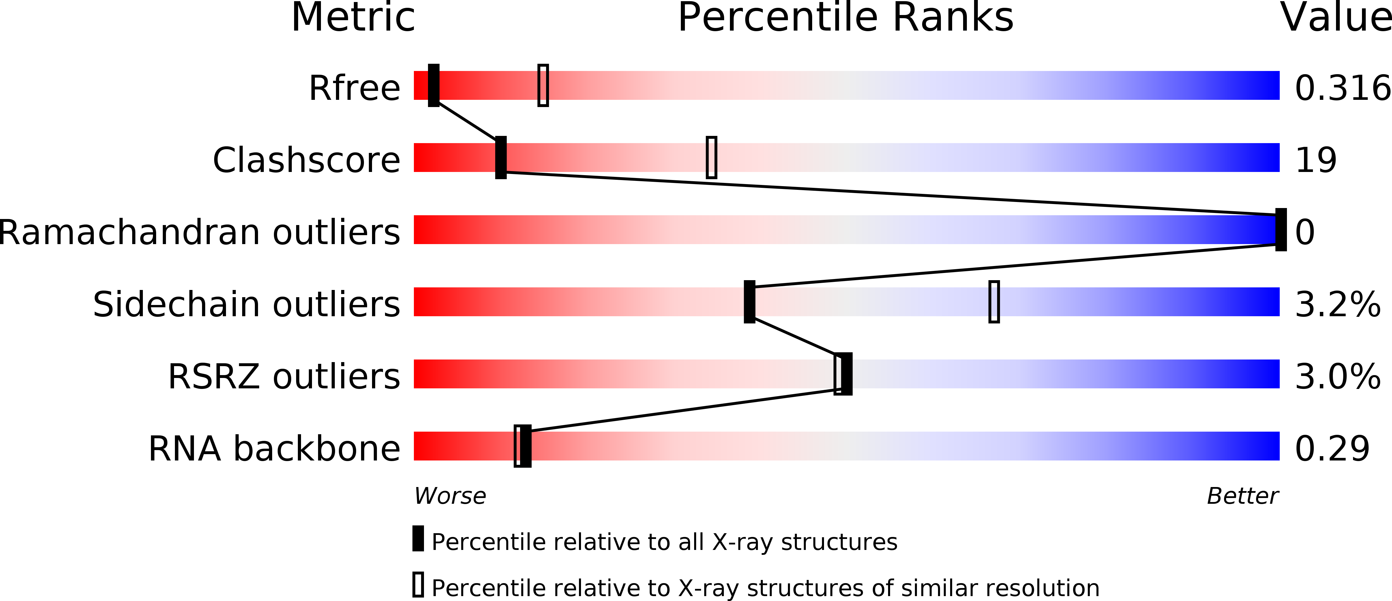
Resolution:
3.40 Å
R-Value Free:
0.31
R-Value Work:
0.26
R-Value Observed:
0.26
Space Group:
P 1 21 1
Macromolecular Entities
Polymer Type:polypeptide(L)
Molecule:50S ribosomal protein L7Ae
Gene (Uniprot):rpl7ae
Chain IDs:A, C
Chain Length:132
Number of Molecules:2
Biological Source:Pyrococcus horikoshii (strain ATCC 700860 / DSM 12428 / JCM 9974 / NBRC 100139 / OT-3)
Polymer Type:polyribonucleotide
Molecule:RNA (47-MER)
Chain IDs:B, D
Chain Length:51
Number of Molecules:2
Biological Source:Pyrococcus horikoshii OT3
Ligand Molecules
Primary Citation
Structural basis for recognition of a kink-turn motif by an archaeal homologue of human RNase P protein Rpp38
Biochem.Biophys.Res.Commun. 474 541 546 (2016)
PMID: 27114305 DOI: 10.1016/j.bbrc.2016.04.118

Abstact

PhoRpp38 in the hyperthermophilic archaeon Pyrococcus horikoshii, a homologue of human ribonuclease P (RNase P) protein Rpp38, belongs to the ribosomal protein L7Ae family that specifically recognizes a kink-turn (K-turn) motif. A previous biochemical study showed that PhoRpp38 specifically binds to two stem-loops, SL12 and SL16, containing helices P12.1/12.2 and P15/16 respectively, in P. horikoshii RNase P RNA (PhopRNA). In order to gain insight into the PhoRpp38 binding mode to PhopRNA, we determined the crystal structure of PhoRpp38 in complex with the SL12 mutant (SL12M) at a resolution of 3.4 Å. The structure revealed that Lys35 on the β-strand (β1) and Asn38, Glu39, and Lys42 on the α-helix (α2) in PhoRpp38 interact with characteristic G•A and A•G pairs in SL12M, where Ile93, Glu94, and Val95, on a loop between α4 and β4 in PhoRpp38, interact with the 3-nucleotide bulge (G-G-U) in the SL12M. The structure demonstrates the previously proposed secondary structure of SL12, including helix P12.2. Structure-based mutational analysis indicated that amino acid residues involved in the binding to SL12 are also responsible for the binding to SL16. This result suggested that each PhoRpp38 binds to the K-turns in SL12 and SL16 in PhopRNA. A pull-down assay further suggested the presence of a second K-turn in SL12. Based on the present results, together with available data, we discuss a structural basis for recognition of K-turn motifs in PhopRNA by PhoRpp38.

Legend

Protein

Chemical

Disease

Primary Citation of related structures