5D81 image
Deposition Date 2015-08-15
Release Date 2015-12-02
Last Version Date 2025-04-02
Entry Detail
PDB ID:
5D81
Keywords:
Title:
Crystal Structure of Ketosteroid Isomerase from Pseudomonas putida (pKSI); D40N, Y57(Cl-Y)
Biological Source:
Source Organism:
Host Organism:
Method Details:
Experimental Method:
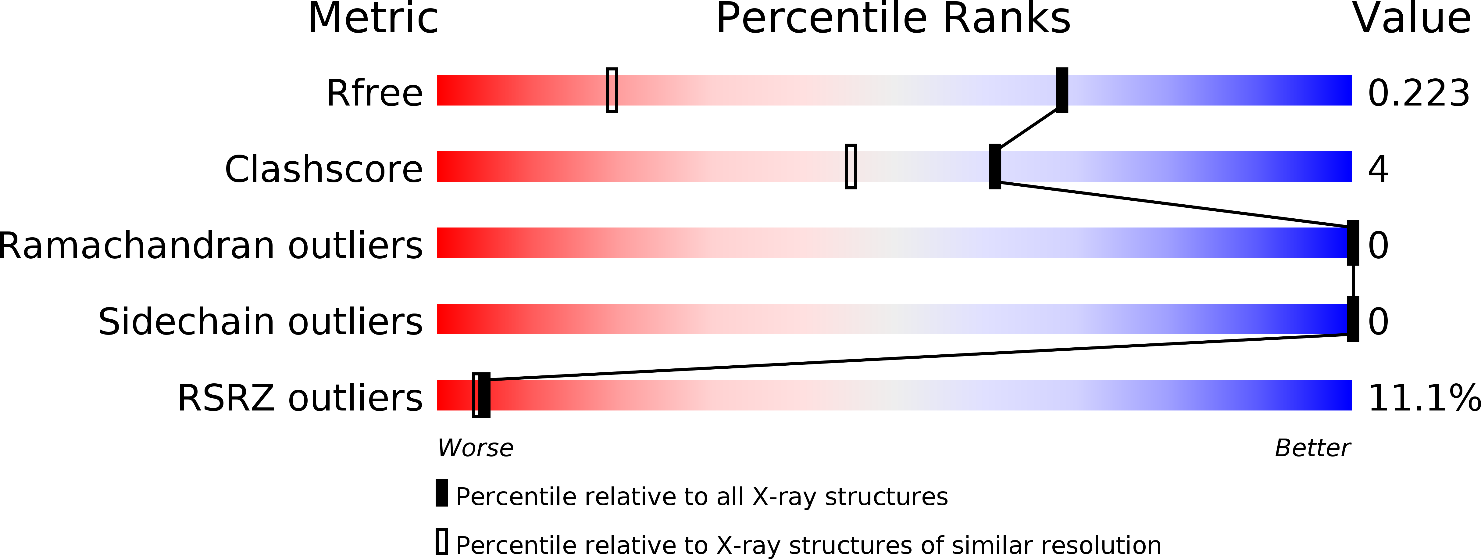
Resolution:
1.39 Å
R-Value Free:
0.22
R-Value Work:
0.18
R-Value Observed:
0.18
Space Group:
C 2 2 21
Macromolecular Entities
Structures with similar UniProt ID
Protein Blast
Polymer Type:polypeptide(L)
Molecule:Delta(5)-3-ketosteroid isomerase
Gene (Uniprot):ksi
Mutagens:D40N,Y57(Cl-Y)
Chain IDs:A
Chain Length:135
Number of Molecules:1
Biological Source:Pseudomonas putida
Ligand Molecules
Primary Citation
Dissecting Proton Delocalization in an Enzyme's Hydrogen Bond Network with Unnatural Amino Acids.
Biochemistry 54 7110 7119 (2015)
PMID: 26571340 DOI: 10.1021/acs.biochem.5b00958

Abstact

Extended hydrogen bond networks are a common structural motif of enzymes. A recent analysis proposed quantum delocalization of protons as a feature present in the hydrogen bond network spanning a triad of tyrosines (Y(16), Y(32), and Y(57)) in the active site of ketosteroid isomerase (KSI), contributing to its unusual acidity and large isotope shift. In this study, we utilized amber suppression to substitute each tyrosine residue with 3-chlorotyrosine to test the delocalization model and the proton affinity balance in the triad. X-ray crystal structures of each variant demonstrated that the structure, notably the O-O distances within the triad, was unaffected by 3-chlorotyrosine substitutions. The changes in the cluster's acidity and the acidity's isotope dependence in these variants were assessed via UV-vis spectroscopy and the proton sharing pattern among individual residues with (13)C nuclear magnetic resonance. Our data show pKa detuning at each triad residue alters the proton delocalization behavior in the H-bond network. The extra stabilization energy necessary for the unusual acidity mainly comes from the strong interactions between Y(57) and Y(16). This is further enabled by Y(32), which maintains the right geometry and matched proton affinity in the triad. This study provides a rich picture of the energetics of the hydrogen bond network in enzymes for further model refinement.

Legend

Protein

Chemical

Disease

Primary Citation of related structures