5D7W image
Deposition Date 2015-08-14
Release Date 2016-01-13
Last Version Date 2023-11-08
Entry Detail
PDB ID:
5D7W
Keywords:
Title:
Crystal structure of serralysin
Biological Source:
Source Organism:
Host Organism:
Method Details:
Experimental Method:
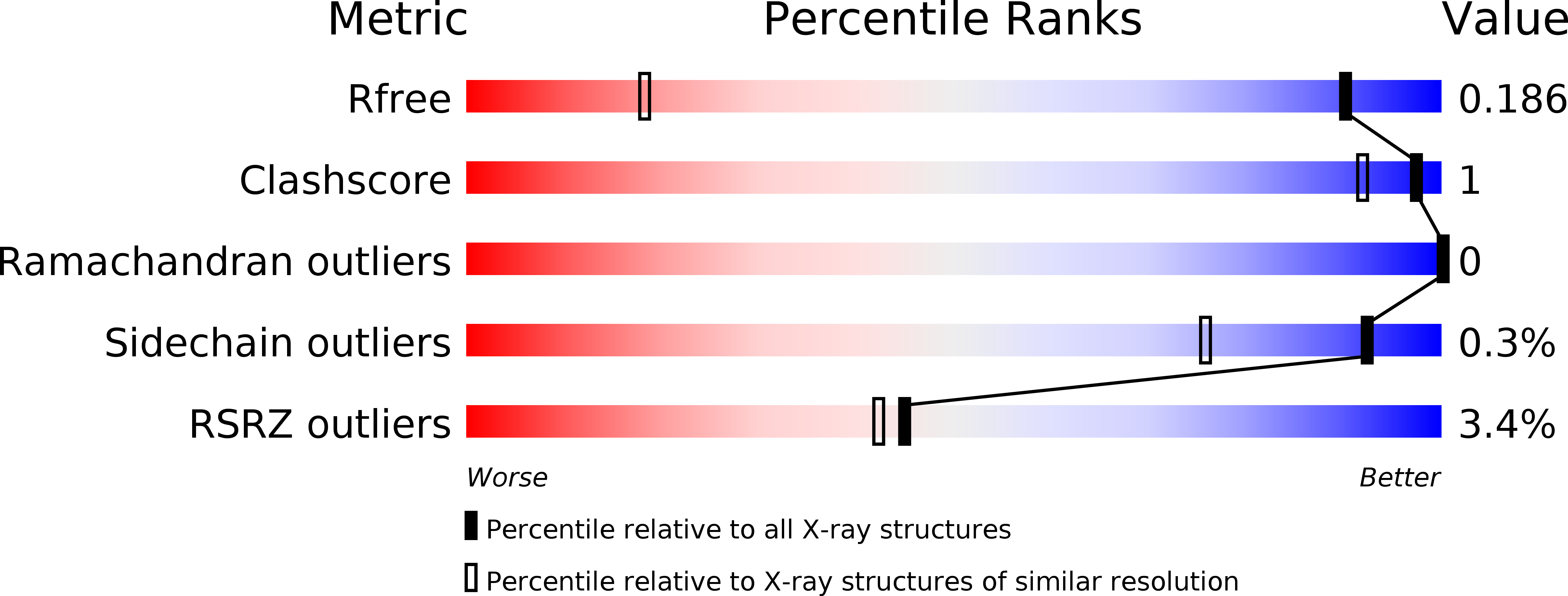
Resolution:
1.10 Å
R-Value Free:
0.18
R-Value Work:
0.17
R-Value Observed:
0.17
Space Group:
P 21 21 21
Macromolecular Entities
Structures with similar UniProt ID
Protein Blast
Polymer Type:polypeptide(L)
Molecule:Serralysin
Mutagens:P250L
Chain IDs:A
Chain Length:469
Number of Molecules:1
Biological Source:Serratia marcescens
Primary Citation
Structure of a thermostable serralysin from Serratia sp. FS14 at 1.1 angstrom resolution.
Acta Crystallogr.,Sect.F 72 10 15 (2016)
PMID: 26750478 DOI: 10.1107/S2053230X15023092

Abstact

Serralysin is a well studied metalloprotease, and typical serralysins are not thermostable. The serralysin isolated from Serratia sp. FS14 was found to be thermostable, and in order to reveal the mechanism responsible for its thermostability, the crystal structure of serralysin from Serratia sp. FS14 was solved to a crystallographic R factor of 0.1619 at 1.10 Å resolution. Similar to its homologues, it mainly consists of two domains: an N-terminal catalytic domain and a `parallel β-roll' C-terminal domain. Comparative studies show that the shape of the catalytic active-site cavity is more open owing to the 189-198 loop, with a short 310-helix protruding further from the molecular surface, and that the β-sheets comprising the `parallel β-roll' are longer than those in its homologues. The formation of hydrogen bonds from one of the nonconserved residues (Asn200) to Lys27 may contribute to the thermostability.

Legend

Protein

Chemical

Disease

Primary Citation of related structures