Deposition Date
2015-08-07
Release Date
2016-03-16
Last Version Date
2024-11-13
Entry Detail
PDB ID:
5D4K
Keywords:
Title:
Crystal structure of the human polymeric Ig receptor (pIgR) ectodomain
Biological Source:
Source Organism(s):
Homo sapiens (Taxon ID: 9606)
Expression System(s):
Method Details:
Experimental Method:
Resolution:
2.60 Å
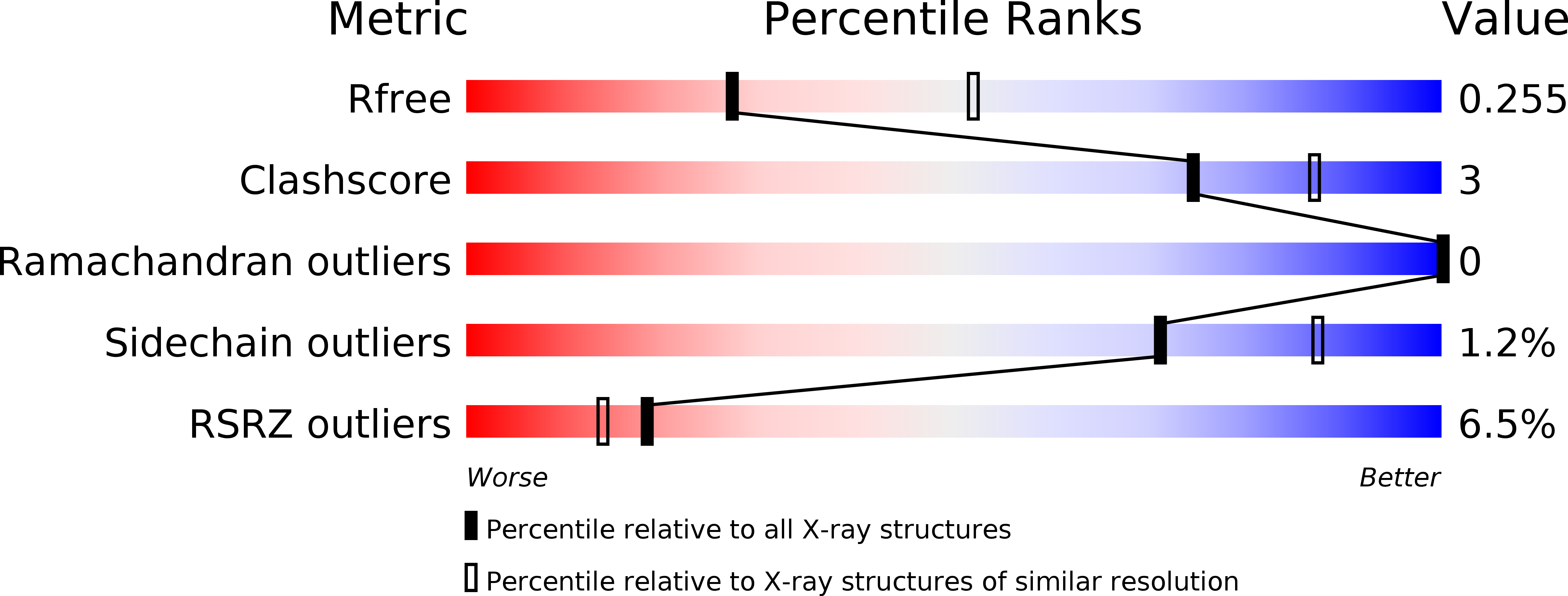
R-Value Free:
0.25
R-Value Work:
0.20
R-Value Observed:
0.20
Space Group:
P 1 21 1