Deposition Date
2015-07-30
Release Date
2015-08-12
Last Version Date
2024-03-13
Entry Detail
PDB ID:
5CYV
Keywords:
Title:
Crystal structure of CouR from Rhodococcus jostii RHA1 bound to p-coumaroyl-CoA
Biological Source:
Source Organism(s):
Rhodococcus jostii (strain RHA1) (Taxon ID: 101510)
Expression System(s):
Method Details:
Experimental Method:
Resolution:
1.52 Å
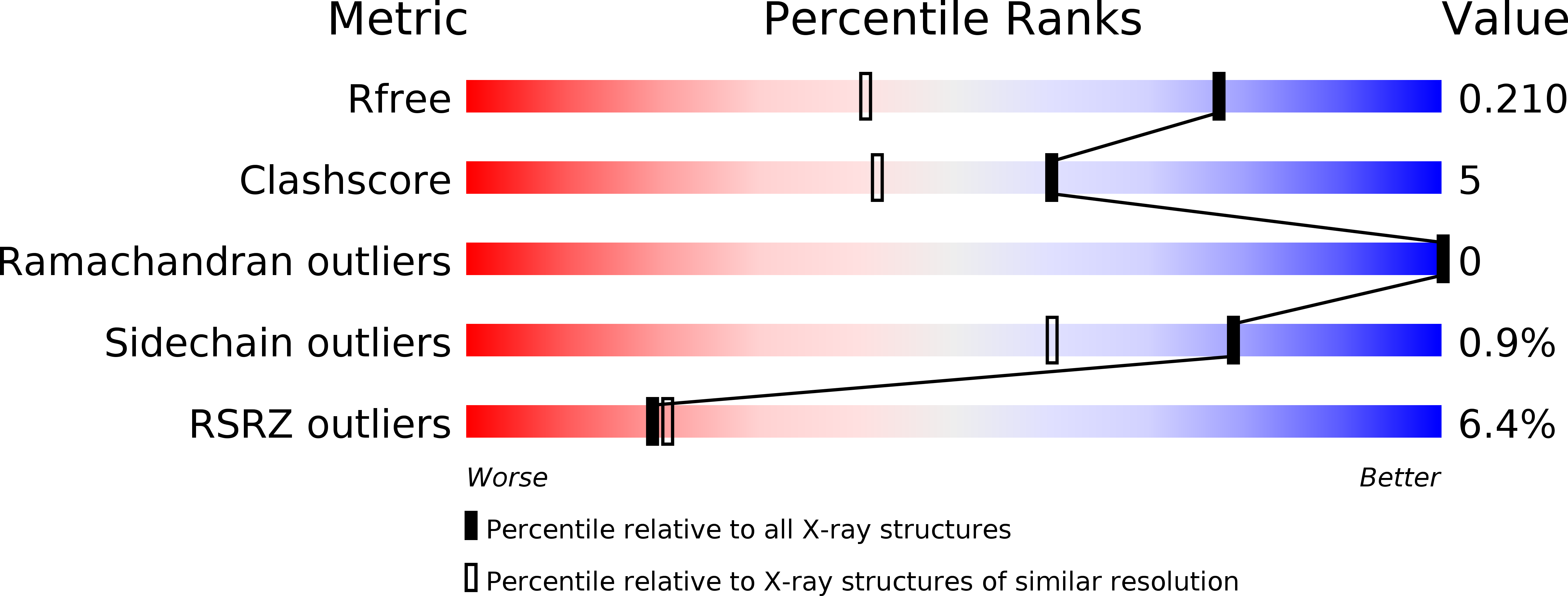
R-Value Free:
0.20
R-Value Work:
0.16
R-Value Observed:
0.16
Space Group:
C 2 2 21