5CUS image
Deposition Date 2015-07-25
Release Date 2015-10-14
Last Version Date 2024-10-16
Entry Detail
PDB ID:
5CUS
Keywords:
Title:
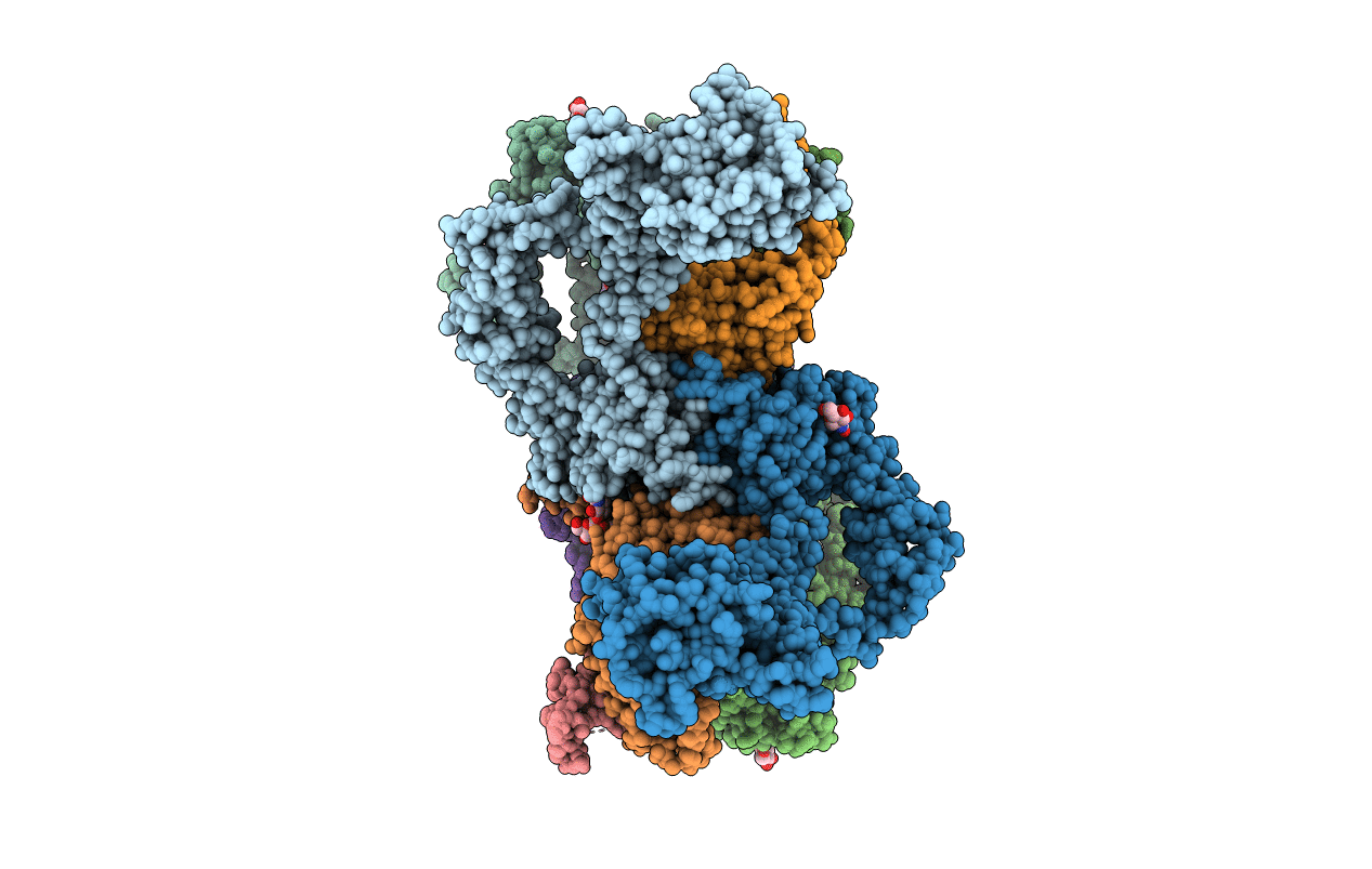
Crystal Structure of sErbB3-Fab3379 Complex
Biological Source:
Source Organism:
Homo sapiens (Taxon ID: 9606)
Host Organism:
Method Details:
Experimental Method:
Resolution:
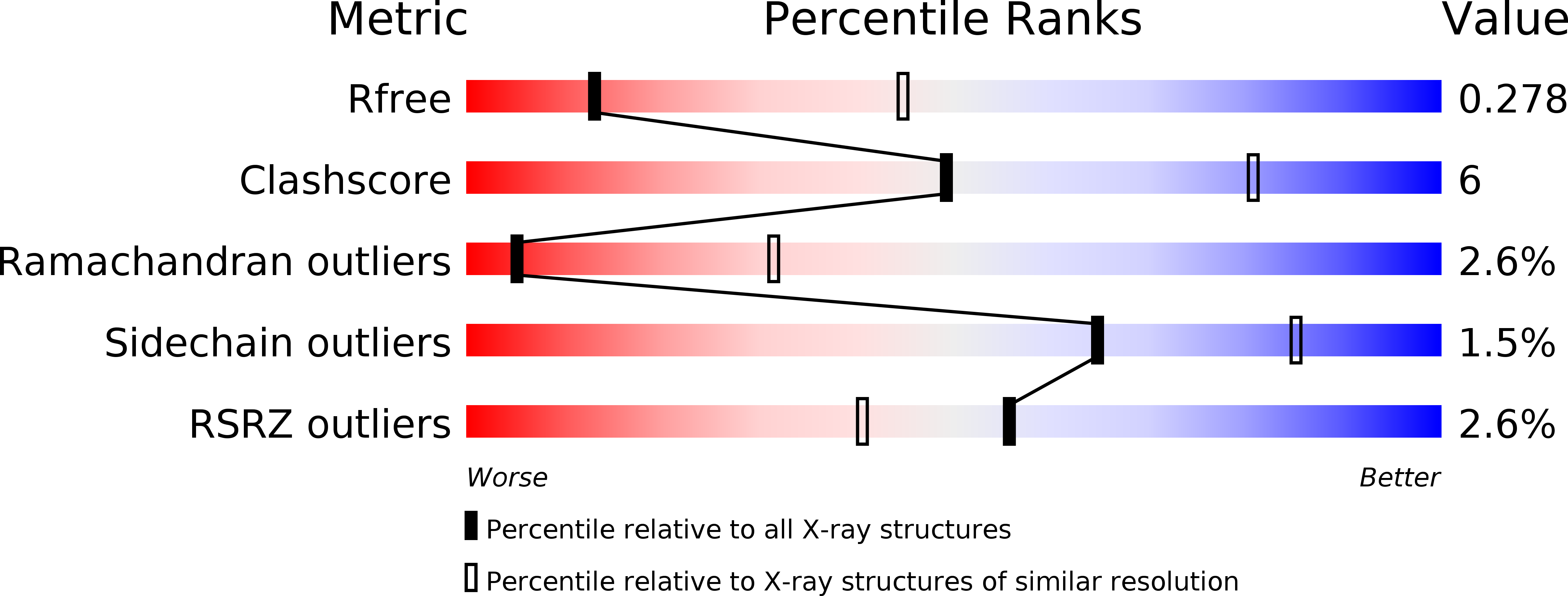
3.20 Å
R-Value Free:
0.27
R-Value Work:
0.25
R-Value Observed:
0.25
Space Group:
P 1
Macromolecular Entities
Polymer Type:polypeptide(L)
Molecule:Receptor tyrosine-protein kinase erbB-3
Gene (Uniprot):ERBB3
Chain IDs:A, B, C, D
Chain Length:627
Number of Molecules:4
Biological Source:Homo sapiens
Polymer Type:polypeptide(L)
Molecule:IgG H chain
Chain IDs:E (auth: H), G (auth: I), H (auth: J), I (auth: K)
Chain Length:221
Number of Molecules:4
Biological Source:Homo sapiens
Polymer Type:polypeptide(L)
Molecule:Fab LC region of KTN3379
Chain IDs:F (auth: L), J (auth: M), K (auth: N), L (auth: O)
Chain Length:216
Number of Molecules:4
Biological Source:Homo sapiens
Ligand Molecules
Primary Citation
Inhibition of ErbB3 by a monoclonal antibody that locks the extracellular domain in an inactive configuration.
Proc.Natl.Acad.Sci.USA 112 13225 13230 (2015)
PMID: 26460020 DOI: 10.1073/pnas.1518361112

Abstact

ErbB3 (HER3) is a member of the EGF receptor (EGFR) family of receptor tyrosine kinases, which, unlike the other three family members, contains a pseudo kinase in place of a tyrosine kinase domain. In cancer, ErbB3 activation is driven by a ligand-dependent mechanism through the formation of heterodimers with EGFR, ErbB2, or ErbB4 or via a ligand-independent process through heterodimerization with ErbB2 overexpressed in breast tumors or other cancers. Here we describe the crystal structure of the Fab fragment of an antagonistic monoclonal antibody KTN3379, currently in clinical development in human cancer patients, in complex with the ErbB3 extracellular domain. The structure reveals a unique allosteric mechanism for inhibition of ligand-dependent or ligand-independent ErbB3-driven cancers by binding to an epitope that locks ErbB3 in an inactive conformation. Given the similarities in the mechanism of ErbB receptor family activation, these findings could facilitate structure-based design of antibodies that inhibit EGFR and ErbB4 by an allosteric mechanism.

Legend

Protein

Chemical

Disease

Primary Citation of related structures