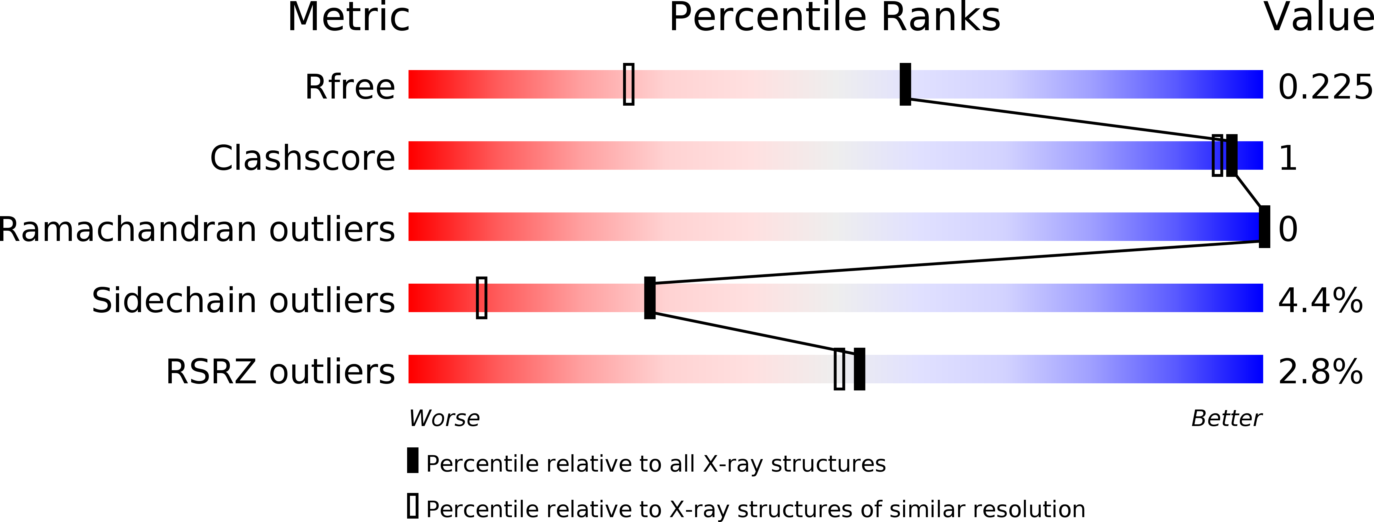
5CRW image
Deposition Date 2015-07-23
Release Date 2015-09-09
Last Version Date 2024-10-16
Entry Detail
PDB ID:
5CRW
Title:
Crystal structure of the b'-a' domain of oxidized protein disulfide isomerase complexed with alpha-synuclein peptide (31-41)
Biological Source:
Source Organism:
Humicola insolens (Taxon ID: 34413)
Homo sapiens (Taxon ID: 9606)
Method Details:
Experimental Method:
Resolution:
1.60 Å
R-Value Free:
0.21
R-Value Work:
0.18
R-Value Observed:
0.18
Space Group:
P 21 21 21
Macromolecular Entities
Structures with similar UniProt ID
Protein Blast
Polymer Type:polypeptide(L)
Molecule:Protein disulfide-isomerase
Chain IDs:A
Chain Length:247
Number of Molecules:1
Biological Source:Humicola insolens
Structures with similar UniProt ID
Protein Blast
Polymer Type:polypeptide(L)
Molecule:Peptide from Alpha-synuclein
Chain IDs:B
Chain Length:11
Number of Molecules:1
Biological Source:Homo sapiens
Primary Citation
Structural basis of redox-dependent substrate binding of protein disulfide isomerase.
Sci Rep 5 13909 13909 (2015)
PMID: 26350503 DOI: 10.1038/srep13909

Abstact

Protein disulfide isomerase (PDI) is a multidomain enzyme, operating as an essential folding catalyst, in which the b' and a' domains provide substrate binding sites and undergo an open-closed domain rearrangement depending on the redox states of the a' domain. Despite the long research history of this enzyme, three-dimensional structural data remain unavailable for its ligand-binding mode. Here we characterize PDI substrate recognition using α-synuclein (αSN) as the model ligand. Our nuclear magnetic resonance (NMR) data revealed that the substrate-binding domains of PDI captured the αSN segment Val37-Val40 only in the oxidized form. Furthermore, we determined the crystal structure of an oxidized form of the b'-a' domains in complex with an undecapeptide corresponding to this segment. The peptide-binding mode observed in the crystal structure with NMR validation, was characterized by hydrophobic interactions on the b' domain in an open conformation. Comparison with the previously reported crystal structure indicates that the a' domain partially masks the binding surface of the b' domain, causing steric hindrance against the peptide in the reduced form of the b'-a' domains that exhibits a closed conformation. These findings provide a structural basis for the mechanism underlying the redox-dependent substrate binding of PDI.

Legend

Protein

Chemical

Disease

Primary Citation of related structures