5CKQ image
Deposition Date 2015-07-15
Release Date 2017-01-18
Last Version Date 2024-10-23
Entry Detail
PDB ID:
5CKQ
Keywords:
Title:
CUB1-EGF-CUB2 domains of rat MASP-1
Biological Source:
Source Organism:
Host Organism:
Method Details:
Experimental Method:
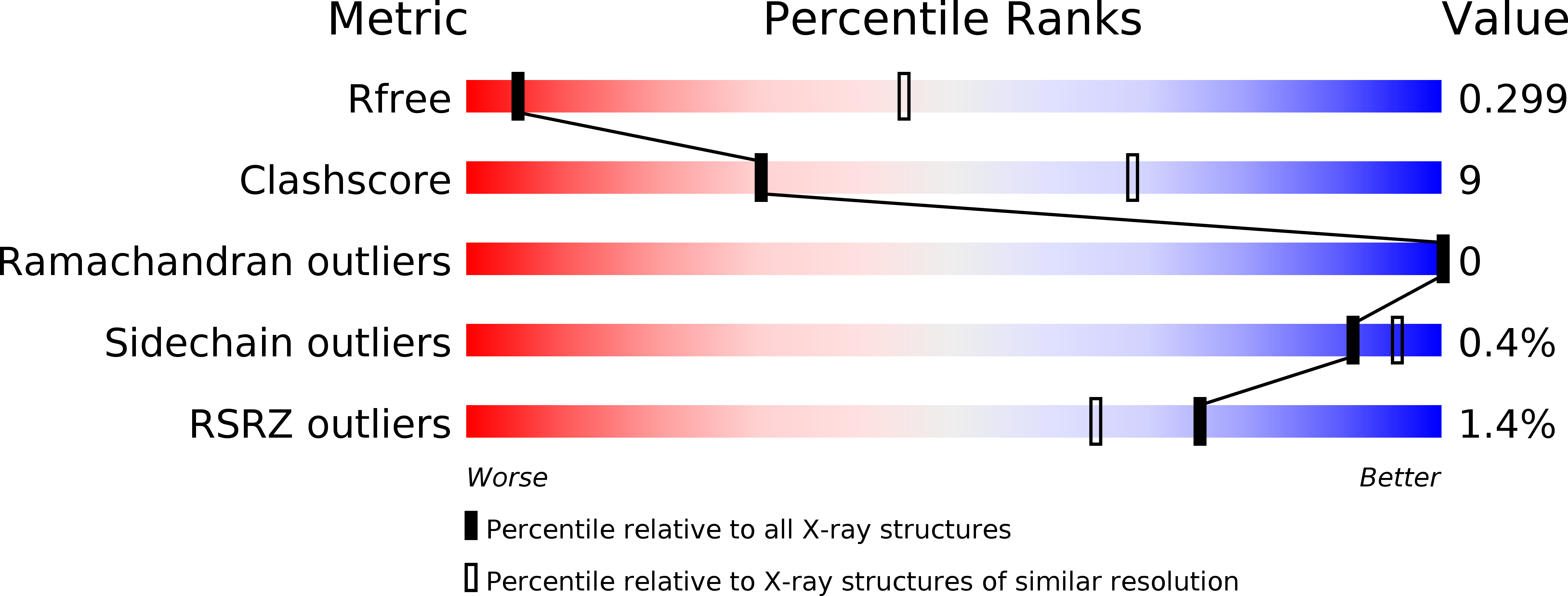
Resolution:
3.70 Å
R-Value Free:
0.29
R-Value Work:
0.24
R-Value Observed:
0.25
Space Group:
I 2 3
Macromolecular Entities
Structures with similar UniProt ID
Protein Blast
Polymer Type:polypeptide(L)
Molecule:Mannan-binding lectin serine protease 1
Gene (Uniprot):Masp1
Chain IDs:A
Chain Length:276
Number of Molecules:1
Biological Source:Rattus norvegicus
Primary Citation
Flexibility in Mannan-Binding Lectin-Associated Serine Proteases-1 and -2 Provides Insight on Lectin Pathway Activation.
Structure 25 364 375 (2017)
PMID: 28111019 DOI: 10.1016/j.str.2016.12.014

Abstact

The lectin pathway of complement is activated by complexes comprising a recognition component (mannose-binding lectin, serum ficolins, collectin-LK or collectin-K1) and a serine protease (MASP-1 or MASP-2). MASP-1 activates MASP-2, and MASP-2 cleaves C4 and C4b-bound C2. To clarify activation, new crystal structures of Ca2+-bound MASP dimers were determined, together with their solution structures from X-ray scattering, analytical ultracentrifugation, and atomistic modeling. Solution structures of the CUB1-EGF-CUB2 dimer of each MASP indicate that the two CUB2 domains were tilted by as much as 90° compared with the crystal structures, indicating considerable flexibility at the EGF-CUB2 junction. Solution structures of the full-length MASP dimers in their zymogen and activated forms revealed similar structures that were much more bent than anticipated from crystal structures. We conclude that MASP-1 and MASP-2 are flexible at multiple sites and that this flexibility may permit both intra- and inter-complex activation.

Legend

Protein

Chemical

Disease

Primary Citation of related structures
Feedback Form
Name
Email
Institute
Feedback