Deposition Date
2015-07-10
Release Date
2016-06-22
Last Version Date
2023-11-15
Entry Detail
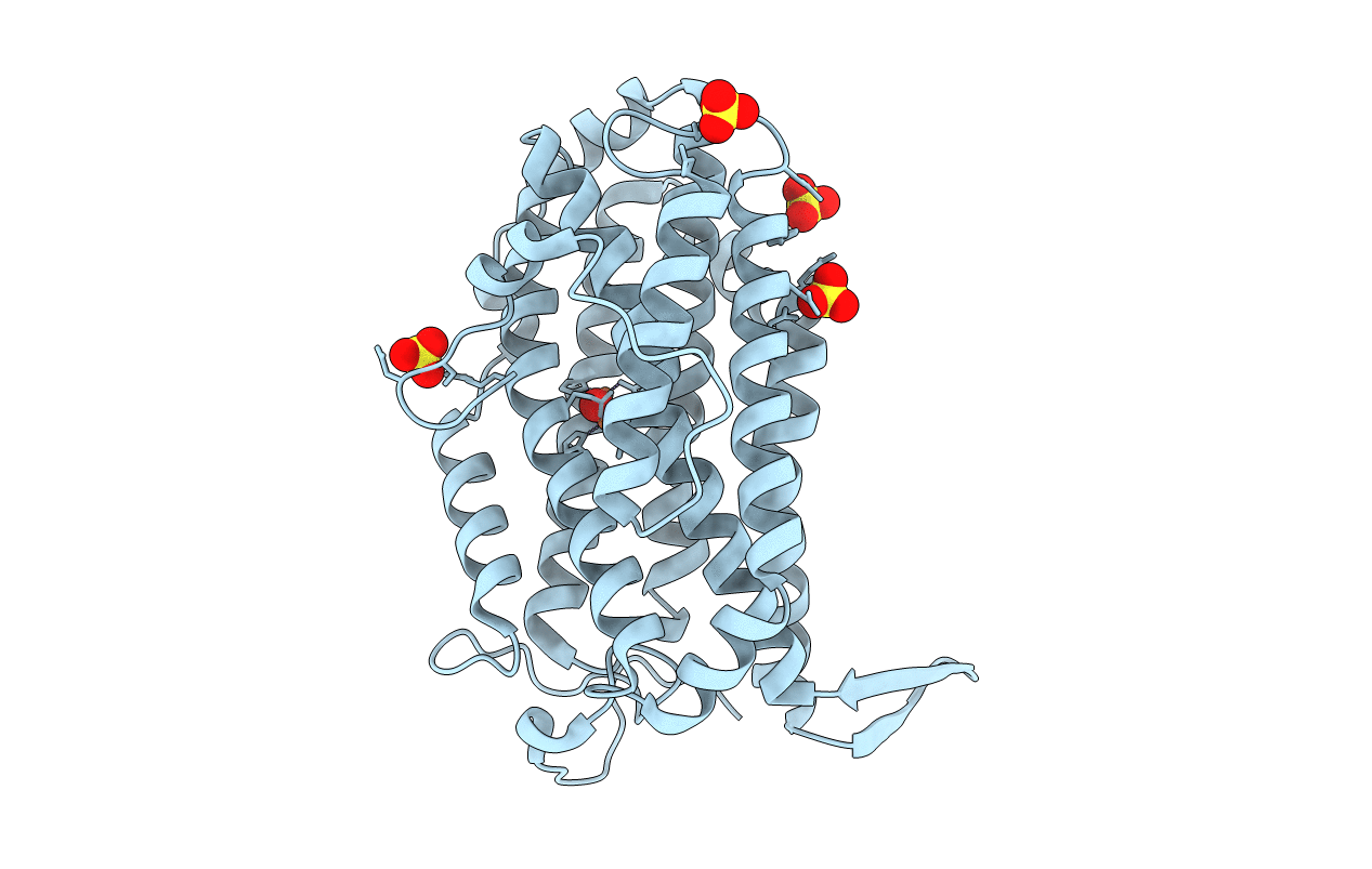
Biological Source:
Source Organism(s):
Escherichia coli 1303 (Taxon ID: 745156)
Expression System(s):
Method Details:
Experimental Method:
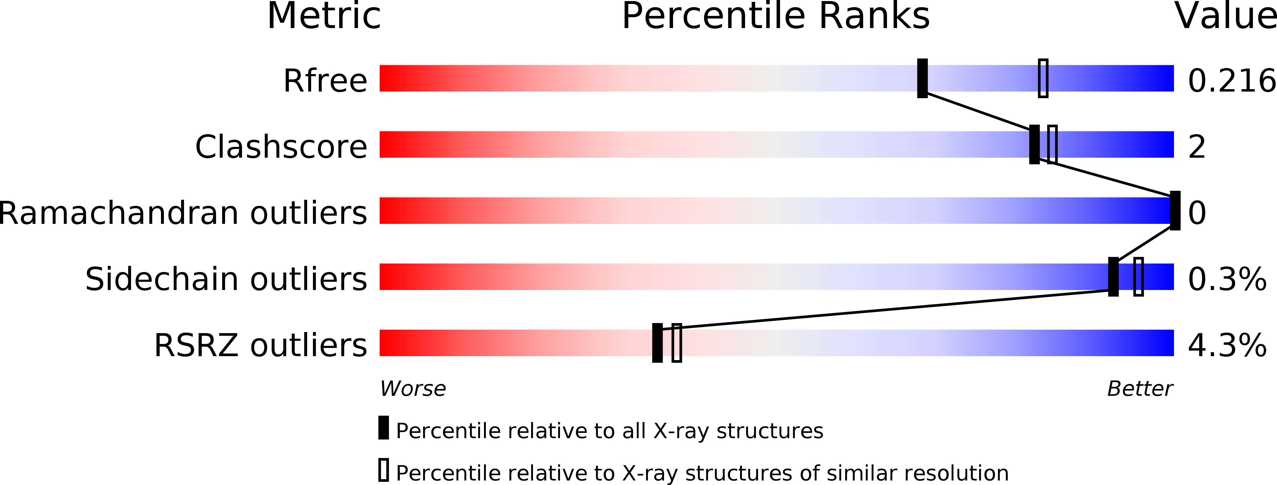
Resolution:
2.25 Å
R-Value Free:
0.21
R-Value Work:
0.18
R-Value Observed:
0.18
Space Group:
P 61 2 2