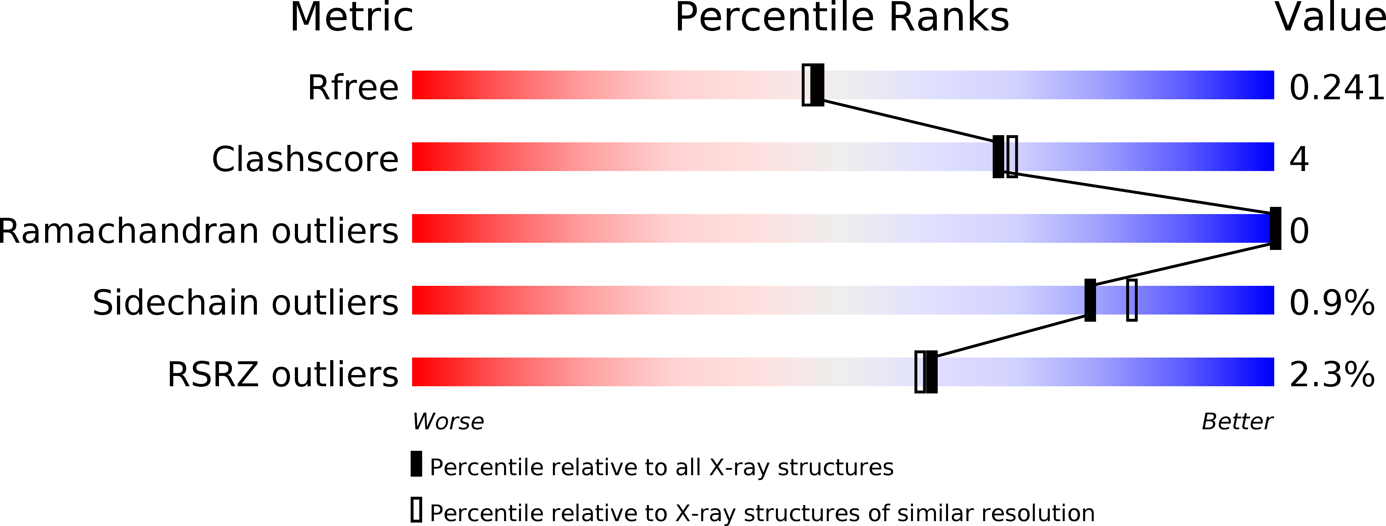
5CBR image
Entry Detail
PDB ID:
5CBR
Title:
Crystal structure of the GluA2 ligand-binding domain (S1S2J) in complex with the antagonist (S)-2-amino-3-(3,4-dichloro-5-(5-hydroxypyridin-3-yl)phenyl)propanoic acid at 2.0A resolution
Biological Source:
Source Organism:
PDB Version:
Deposition Date:
2015-07-01
Release Date:
2015-12-30
Method Details:
Experimental Method:
Resolution:
2.00 Å
R-Value Free:
0.23
R-Value Work:
0.18
R-Value Observed:
0.18
Space Group:
P 21 21 2
Macromolecular Entities
Polymer Type:polypeptide(L)
Description:Glutamate receptor 2,Glutamate receptor 2
Chain IDs:A
Chain Length:264
Number of Molecules:1
Biological Source:Rattus norvegicus
Primary Citation
Studies on Aryl-Substituted Phenylalanines: Synthesis, Activity, and Different Binding Modes at AMPA Receptors.
J.Med.Chem. 59 448 461 (2016)
PMID: 26653877 DOI: 10.1021/acs.jmedchem.5b01666

Abstact

A series of racemic aryl-substituted phenylalanines was synthesized and evaluated in vitro at recombinant rat GluA1-3, at GluK1-3, and at native AMPA receptors. The individual enantiomers of two target compounds, (RS)-2-amino-3-(3,4-dichloro-5-(5-hydroxypyridin-3-yl)phenyl)propanoic acid 37 and (RS)-2-amino-3-(3'-hydroxybiphenyl-3-yl)propanoic acid 38, were characterized. (S)-37 and (R)-38 were identified as the only biologically active isomers, both being antagonists at GluA2 receptors with Kb of 1.80 and 3.90 μM, respectively. To address this difference in enantiopharmacology, not previously seen for amino acid-based AMPA receptor antagonists, X-ray crystal structures of both eutomers in complex with the GluA2 ligand binding domain were solved. The cocrystal structures of (S)-37 and (R)-38 showed similar interactions of the amino acid parts but unexpected and different orientations and interactions of the biaromatic parts of the ligands inside the binding site, with (R)-38 having a binding mode not previously identified for amino acid-based antagonists.

Legend

Protein

Chemical

Disease

Primary Citation of related structures