Deposition Date
2015-06-30
Release Date
2016-02-10
Last Version Date
2023-11-08
Entry Detail
PDB ID:
5CB0
Keywords:
Title:
Crystal structure and functional implications of the tandem-type universal stress protein UspE from Escherichia coli
Biological Source:
Source Organism(s):
Escherichia coli (Taxon ID: 83333)
Expression System(s):
Method Details:
Experimental Method:
Resolution:
3.21 Å
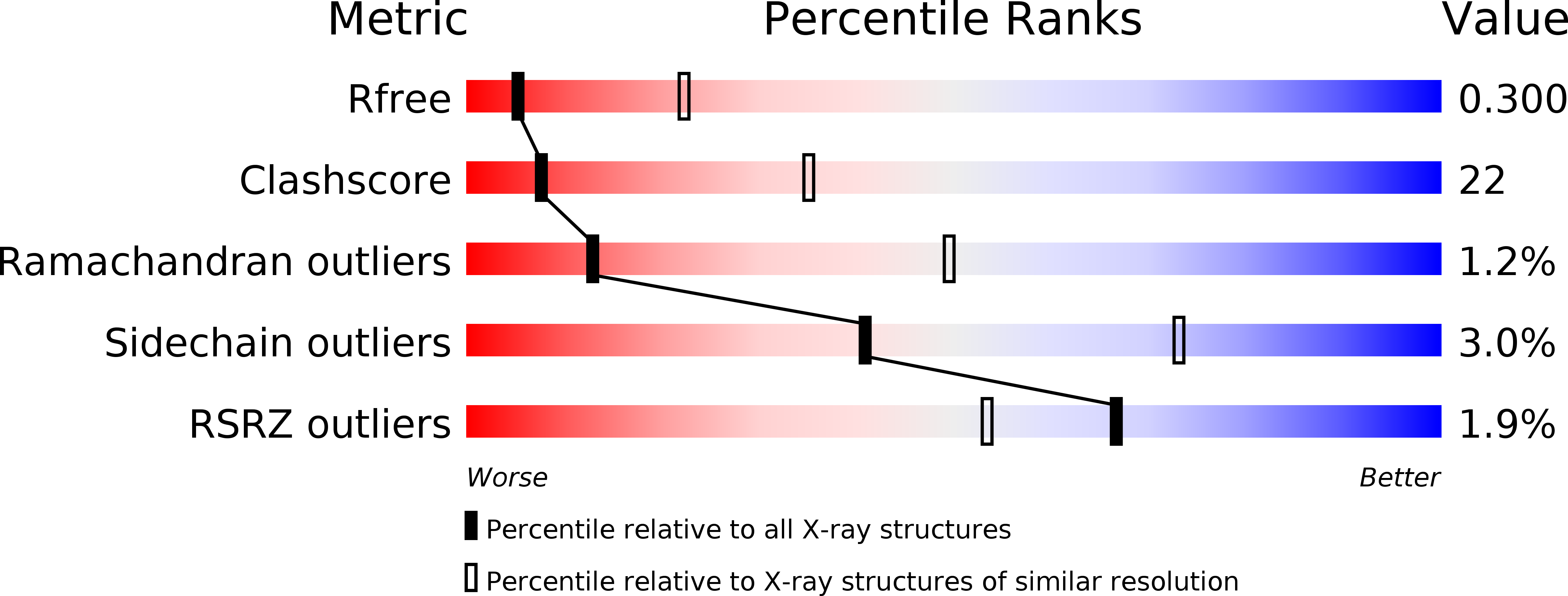
R-Value Free:
0.30
R-Value Work:
0.24
R-Value Observed:
0.24
Space Group:
I 41 2 2