Deposition Date
2015-06-29
Release Date
2015-09-09
Last Version Date
2024-11-06
Entry Detail
PDB ID:
5CAM
Keywords:
Title:
Crystal Structure of the Cytoplasmic Domain of the Pseudomonas putida Anti-sigma Factor PupR (SeMet)
Biological Source:
Source Organism(s):
Pseudomonas putida (Taxon ID: 303)
Expression System(s):
Method Details:
Experimental Method:
Resolution:
2.17 Å
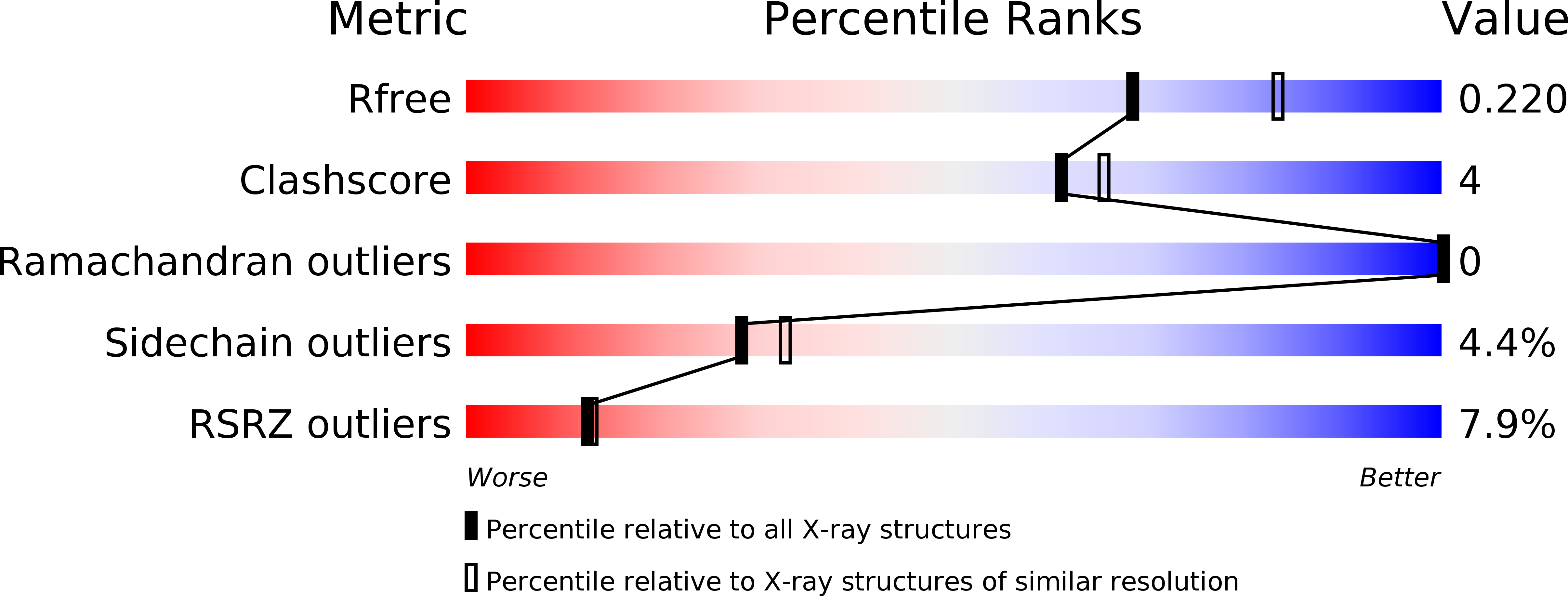
R-Value Free:
0.21
R-Value Work:
0.17
R-Value Observed:
0.18
Space Group:
P 1 21 1