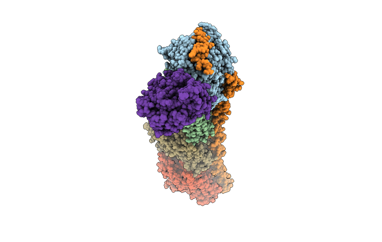
5CA0 image
Entry Detail
PDB ID:
5CA0
Title:
Crystal structure of T2R-TTL-Lexibulin complex
Biological Source:
Host Organism:
PDB Version:
Deposition Date:
2015-06-29
Release Date:
2015-11-04
Method Details:
Experimental Method:
Resolution:
2.50 Å
R-Value Free:
0.23
R-Value Work:
0.19
R-Value Observed:
0.19
Space Group:
P 21 21 21
Macromolecular Entities
Polymer Type:polypeptide(L)
Description:Tubulin alpha-1B chain
Chain IDs:A, C
Chain Length:451
Number of Molecules:2
Biological Source:Sus scrofa
Polymer Type:polypeptide(L)
Description:Uncharacterized protein
Chain IDs:B, D
Chain Length:445
Number of Molecules:2
Biological Source:Sus scrofa
Polymer Type:polypeptide(L)
Description:Stathmin-4
Chain IDs:E
Chain Length:143
Number of Molecules:1
Biological Source:Rattus norvegicus
Polymer Type:polypeptide(L)
Description:Uncharacterized protein
Chain IDs:F
Chain Length:384
Number of Molecules:1
Biological Source:Gallus gallus
Primary Citation
Structures of a diverse set of colchicine binding site inhibitors in complex with tubulin provide a rationale for drug discovery.
Febs J. 283 102 111 (2016)
PMID: 26462166 DOI: 10.1111/febs.13555

Abstact

UNLABELLED Microtubules are dynamic assemblies of αβ-tubulin heterodimers and have been recognized as highly attractive targets for cancer chemotherapy. A broad range of agents bind to tubulin and interfere with microtubule assembly. Despite having a long history of characterization, colchicine binding site inhibitors (CBSIs) have not yet reached the commercial phase as anti-cancer drugs to date. We determined the structures of tubulin complexed with a set of structurally diverse CBSIs (lexibulin, nocodazole, plinabulin and tivantinib), among which nocodazole and tivantinib are both binary-function inhibitors targeting cancer-related kinases and microtubules simultaneously. High resolution structures revealed the detailed interactions between these ligands and tubulin. Our results showed that the binding modes of the CBSIs were different from previous docking models, highlighting the importance of crystal structure information in structure-based drug design. A real structure-based pharmacophore was proposed to rationalize key common interactions of the CBSIs at the colchicine domain. Our studies provide a solid structural basis for developing new anti-cancer agents for the colchicine binding site. DATABASE The atomic coordinates and structure factors for tubulin complexed with lexibulin, nocodazole, plinabulin and tivantinib have been deposited in the Protein Data Bank under accession codes 5CA0, 5CA1, 5C8Y and 5CB4, respectively.

Legend

Protein

Chemical

Disease

Primary Citation of related structures