5C7R image
Deposition Date 2015-06-24
Release Date 2015-09-30
Last Version Date 2023-09-27
Entry Detail
PDB ID:
5C7R
Title:
Revealing surface waters on an antifreeze protein by fusion protein crystallography
Biological Source:
Source Organism:
Host Organism:
Method Details:
Experimental Method:
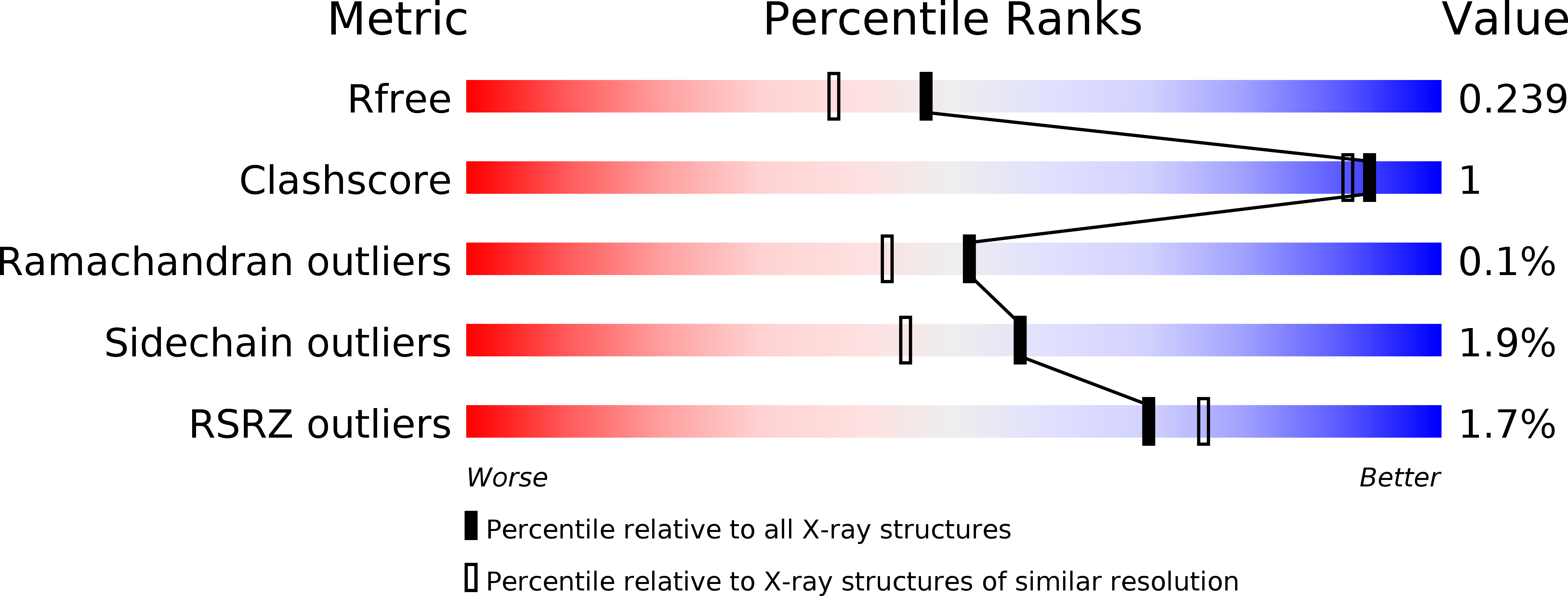
Resolution:
1.94 Å
R-Value Free:
0.23
R-Value Work:
0.17
R-Value Observed:
0.17
Space Group:
P 1 21 1
Macromolecular Entities
Polymer Type:polypeptide(L)
Molecule:Fusion protein of Maltose-binding periplasmic protein and Type-3 ice-structuring protein HPLC 12
Chain IDs:A, B
Chain Length:444
Number of Molecules:2
Biological Source:Escherichia coli O157:H7, Zoarces americanus
Ligand Molecules
Peptide-like Molecules
PRD_900009
Primary Citation
Revealing Surface Waters on an Antifreeze Protein by Fusion Protein Crystallography Combined with Molecular Dynamic Simulations.
J.Phys.Chem.B 119 12808 12815 (2015)
PMID: 26371748 DOI: 10.1021/acs.jpcb.5b06474

Abstact

Antifreeze proteins (AFPs) adsorb to ice through an extensive, flat, relatively hydrophobic surface. It has been suggested that this ice-binding site (IBS) organizes surface waters into an ice-like clathrate arrangement that matches and fuses to the quasi-liquid layer on the ice surface. On cooling, these waters join the ice lattice and freeze the AFP to its ligand. Evidence for the generality of this binding mechanism is limited because AFPs tend to crystallize with their IBS as a preferred protein-protein contact surface, which displaces some bound waters. Type III AFP is a 7 kDa globular protein with an IBS made up two adjacent surfaces. In the crystal structure of the most active isoform (QAE1), the part of the IBS that docks to the primary prism plane of ice is partially exposed to solvent and has clathrate waters present that match this plane of ice. The adjacent IBS, which matches the pyramidal plane of ice, is involved in protein-protein crystal contacts with few surface waters. Here we have changed the protein-protein contacts in the ice-binding region by crystallizing a fusion of QAE1 to maltose-binding protein. In this 1.9 Å structure, the IBS that fits the pyramidal plane of ice is exposed to solvent. By combining crystallography data with MD simulations, the surface waters on both sides of the IBS were revealed and match well with the target ice planes. The waters on the pyramidal plane IBS were loosely constrained, which might explain why other isoforms of type III AFP that lack the prism plane IBS are less active than QAE1. The AFP fusion crystallization method can potentially be used to force the exposure to solvent of the IBS on other AFPs to reveal the locations of key surface waters.

Legend

Protein

Chemical

Disease

Primary Citation of related structures