Deposition Date
2015-06-24
Release Date
2016-01-20
Last Version Date
2024-03-06
Entry Detail
PDB ID:
5C7Q
Keywords:
Title:
Crystal Structure of the Bdellovibrio bacteriovorus Nucleoside Diphosphate Sugar Hydrolase
Biological Source:
Source Organism(s):
Bdellovibrio bacteriovorus (Taxon ID: 264462)
Expression System(s):
Method Details:
Experimental Method:
Resolution:
1.52 Å
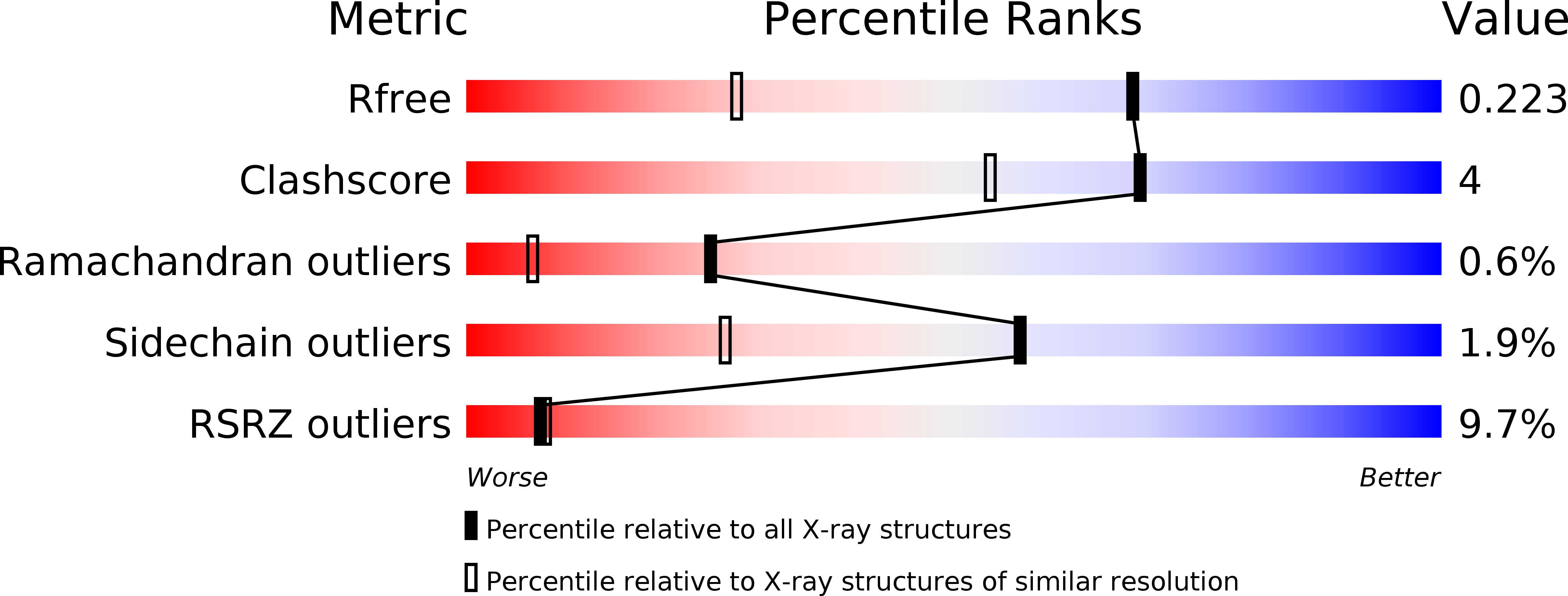
R-Value Free:
0.21
R-Value Work:
0.18
R-Value Observed:
0.18
Space Group:
P 21 21 2