
Deposition Date
2015-06-24
Release Date
2015-10-07
Last Version Date
2024-03-06
Entry Detail
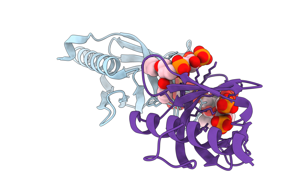
PDB ID:
5C79
Keywords:
Title:
PH domain of ASAP1 in complex with diC4-PtdIns(4,5)P2
Biological Source:
Source Organism(s):
Mus musculus (Taxon ID: 10090)
Expression System(s):
Method Details:
Experimental Method:
Resolution:
1.60 Å
R-Value Free:
0.23
R-Value Work:
0.19
R-Value Observed:
0.20
Space Group:
P 1 21 1
