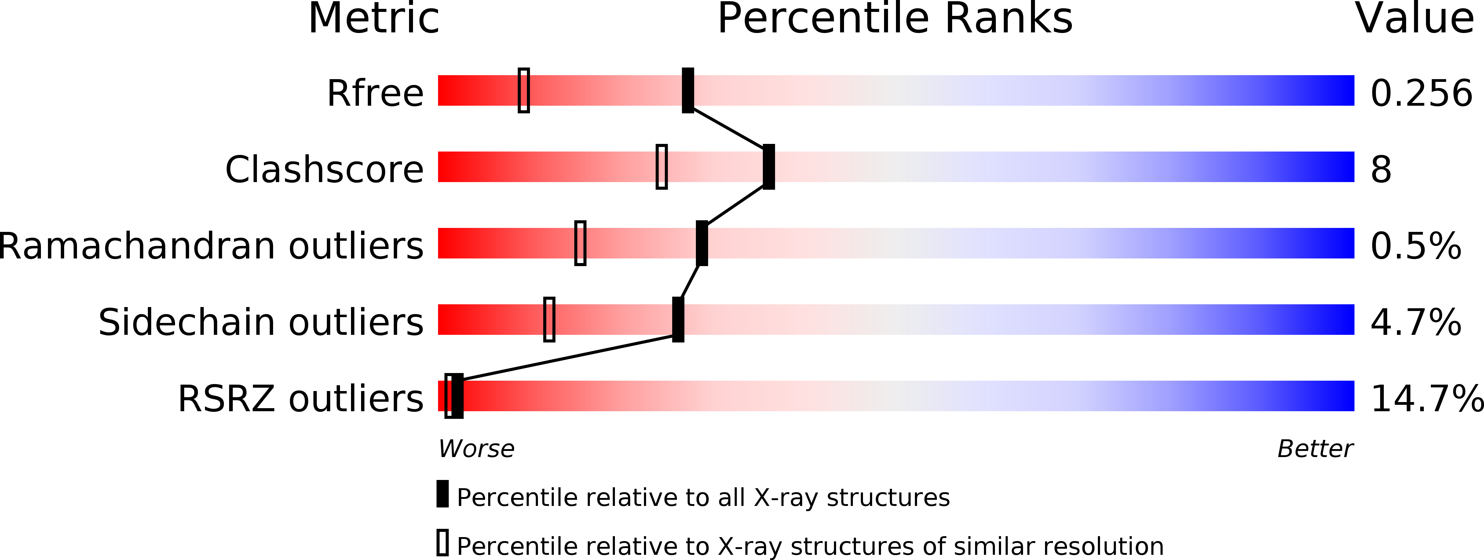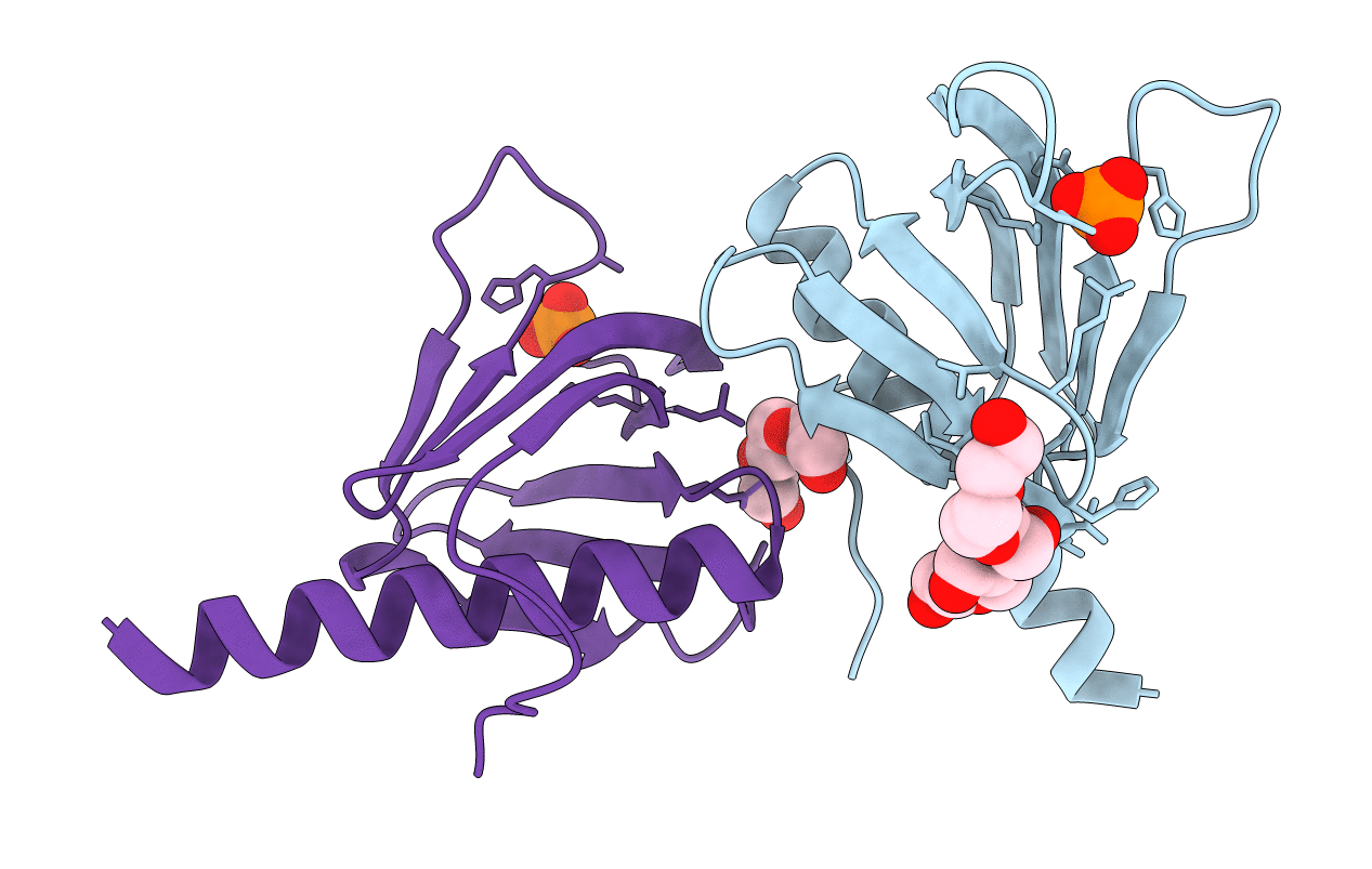
5C6R image
Deposition Date 2015-06-23
Release Date 2015-10-07
Last Version Date 2023-09-27
Entry Detail
PDB ID:
5C6R
Title:
Crystal structure of PH domain of ASAP1
Biological Source:
Source Organism:
Mus musculus (Taxon ID: 10090)
Host Organism:
Method Details:
Experimental Method:
Resolution:
1.80 Å
R-Value Free:
0.25
R-Value Work:
0.20
R-Value Observed:
0.21
Space Group:
P 43
Macromolecular Entities
Polymer Type:polypeptide(L)
Molecule:Arf-GAP
Gene (Uniprot):Asap1
Chain IDs:A, B
Chain Length:150
Number of Molecules:2
Biological Source:Mus musculus
Primary Citation
Molecular Basis for Cooperative Binding of Anionic Phospholipids to the PH Domain of the Arf GAP ASAP1.
Structure 23 1977 1988 (2015)
PMID: 26365802 DOI: 10.1016/j.str.2015.08.008

Abstact

We have defined the molecular basis for association of the PH domain of the Arf GAP ASAP1 with phospholipid bilayers. Structures of the unliganded and dibutyryl PtdIns(4,5)P2-bound PH domain were solved. PtdIns(4,5)P2 made contact with both a canonical site (C site) and an atypical site (A site). We hypothesized cooperative binding of PtdIns(4,5)P2 to the C site and a nonspecific anionic phospholipid to the A site. PtdIns(4,5)P2 dependence of binding to large unilamellar vesicles and GAP activity was sigmoidal, consistent with cooperative sites. In contrast, PtdIns(4,5)P2 binding to the PH domain of PLC δ1 was hyperbolic. Mutation of amino acids in either the C or A site resulted in decreased PtdIns(4,5)P2-dependent binding to vesicles and decreased GAP activity. The results support the idea of cooperative phospholipid binding to the C and A sites of the PH domain of ASAP1. We propose that the mechanism underlies rapid switching between active and inactive ASAP1.

Legend

Protein

Chemical

Disease

Primary Citation of related structures