5C5E image
Deposition Date 2015-06-19
Release Date 2015-08-05
Last Version Date 2023-09-27
Entry Detail
PDB ID:
5C5E
Keywords:
Title:
Structure of KaiA dimer in complex with C-terminal KaiC peptide at 2.8 A resolution
Biological Source:
Host Organism:
Method Details:
Experimental Method:
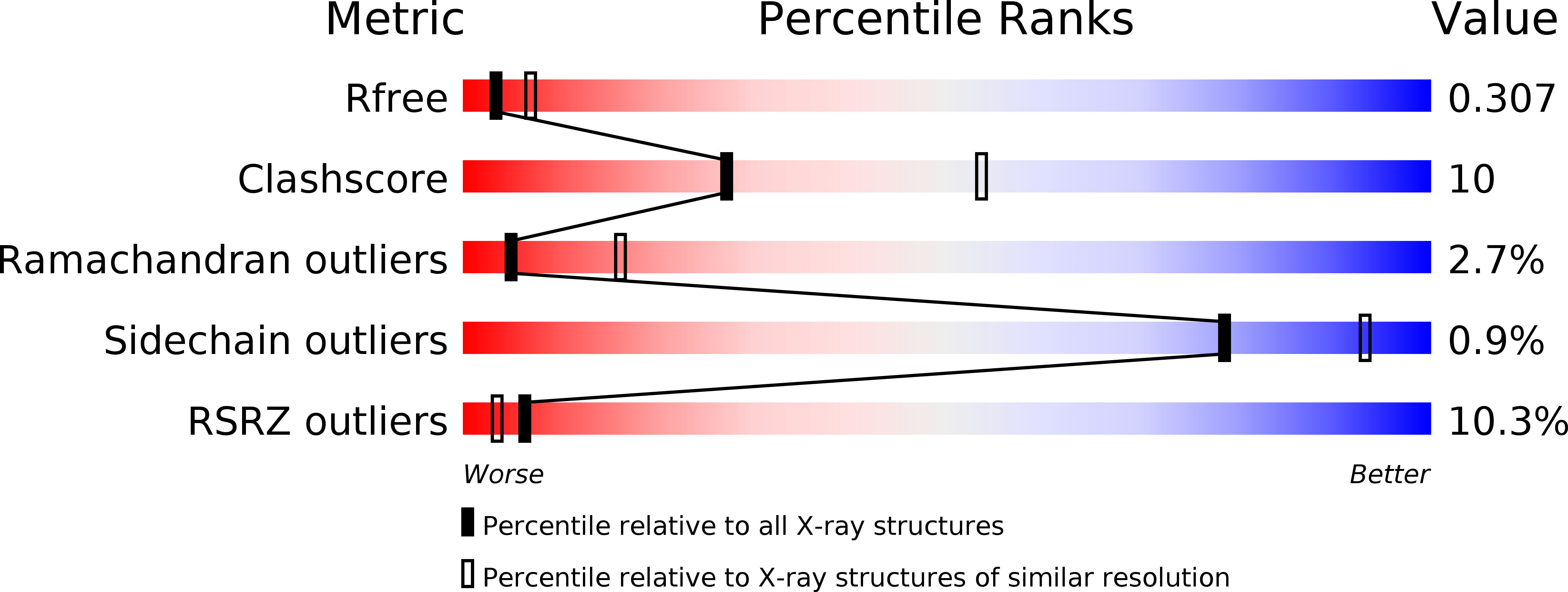
Resolution:
2.82 Å
R-Value Free:
0.30
R-Value Work:
0.23
R-Value Observed:
0.24
Space Group:
P 43 21 2
Macromolecular Entities
Polymer Type:polypeptide(L)
Molecule:Circadian clock protein KaiA
Gene (Uniprot):kaiA
Chain IDs:A, C (auth: B)
Chain Length:290
Number of Molecules:2
Biological Source:Synechococcus elongatus (strain PCC 7942)
Polymer Type:polypeptide(L)
Molecule:KaiC C-terminal peptide
Chain IDs:B (auth: G), D (auth: H)
Chain Length:20
Number of Molecules:2
Biological Source:synthetic construct
Ligand Molecules
Primary Citation
Protein-Protein Interactions in the Cyanobacterial Circadian Clock: Structure of KaiA Dimer in Complex with C-Terminal KaiC Peptides at 2.8 angstrom Resolution.
Biochemistry 54 4575 4578 (2015)
PMID: 26200123 DOI: 10.1021/acs.biochem.5b00694

Abstact

In the cyanobacterial circadian clock, the KaiA, -B, and -C proteins with ATP constitute a post-translational oscillator. KaiA stimulates the KaiC autokinase, and KaiB antagonizes KaiA action. KaiA contacts the intrinsically disordered C-terminal regions of KaiC hexamer to promote phosphorylation across subunit interfaces. The crystal structure of KaiA dimer from Synechococcus elongatus with two KaiC C-terminal 20mer peptides bound reveals that the latter adopt an α-helical conformation and contact KaiA α-helical bundles via mostly hydrophobic interactions. This complex and the crystal structure of KaiC hexamer with truncated C-terminal tails can be fit into the electron microscopy (EM) density of the KaiA:KaiC complex. The hybrid model helps rationalize clock phenotypes of KaiA and KaiC mutants.

Legend

Protein

Chemical

Disease

Primary Citation of related structures