5C3Z image
Deposition Date 2015-06-17
Release Date 2016-06-15
Last Version Date 2025-10-29
Entry Detail
PDB ID:
5C3Z
Keywords:
Title:
Crystal structure of human ribokinase in complex with AMPPCP in C2 spacegroup
Biological Source:
Source Organism:
Homo sapiens (Taxon ID: 9606)
Method Details:
Experimental Method:
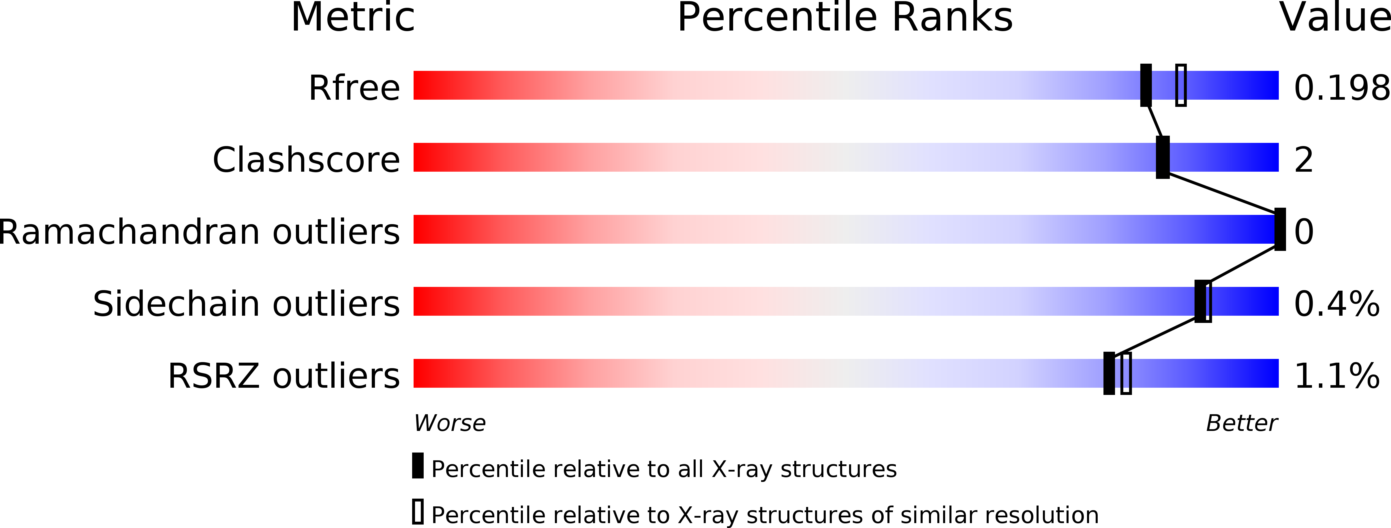
Resolution:
1.90 Å
R-Value Free:
0.18
R-Value Work:
0.14
R-Value Observed:
0.15
Space Group:
C 1 2 1
Macromolecular Entities
Structures with similar UniProt ID
Protein Blast
Polymer Type:polypeptide(L)
Molecule:Ribokinase
Gene (Uniprot):RBKS
Chain IDs:A, B
Chain Length:330
Number of Molecules:2
Biological Source:Homo sapiens
Primary Citation
Conformational flexibility of human ribokinase captured in seven crystal structures.
Int.J.Biol.Macromol. 299 140109 140109 (2025)
PMID: 39837438 DOI: 10.1016/j.ijbiomac.2025.140109

Abstact

d-ribose is a critical sugar substrate involved in the biosynthesis of nucleotides, amino acids, and cofactors, with its phosphorylation to ribose-5-phosphate by ribokinase (RK) constituting the initial step in its metabolism. RK is conserved across all domains of life, and its activity is significantly enhanced by monovalent metal (M+) ions, particularly K+, although the precise mechanism of this activation remains unclear. In this study, we present several crystal structures of human RK in both unliganded and substrate-bound states, offering detailed insights into its substrate binding process, reaction mechanism, and conformational changes throughout the catalytic cycle. Notably, bound ATP exhibited significant conformational flexibility in its triphosphate moiety, a feature shared with other RK homologues, suggesting that achieving a catalytically productive triphosphate configuration plays a key role in regulating enzyme activity. We also identified a unique conformational change in the M+ ion binding loop of human RK, specifically the flipping of the Gly306-Thr307 peptide plane, likely influenced by the ionic radius of the bound ion. These findings provide new insights into the RK reaction mechanism and its activation by M+ ions, paving the way for future investigations into the allosteric regulation of human RK and related sugar kinase enzymes.

Legend

Protein

Chemical

Disease

Primary Citation of related structures
Feedback Form
Name
Email
Institute
Feedback