Deposition Date
2015-06-16
Release Date
2015-08-05
Last Version Date
2024-03-06
Entry Detail
PDB ID:
5C30
Keywords:
Title:
Crystal Structure of Rabbit Ryanodine Receptor 1 Repeat12 Domain
Biological Source:
Source Organism(s):
Oryctolagus cuniculus (Taxon ID: 9986)
Expression System(s):
Method Details:
Experimental Method:
Resolution:
1.55 Å
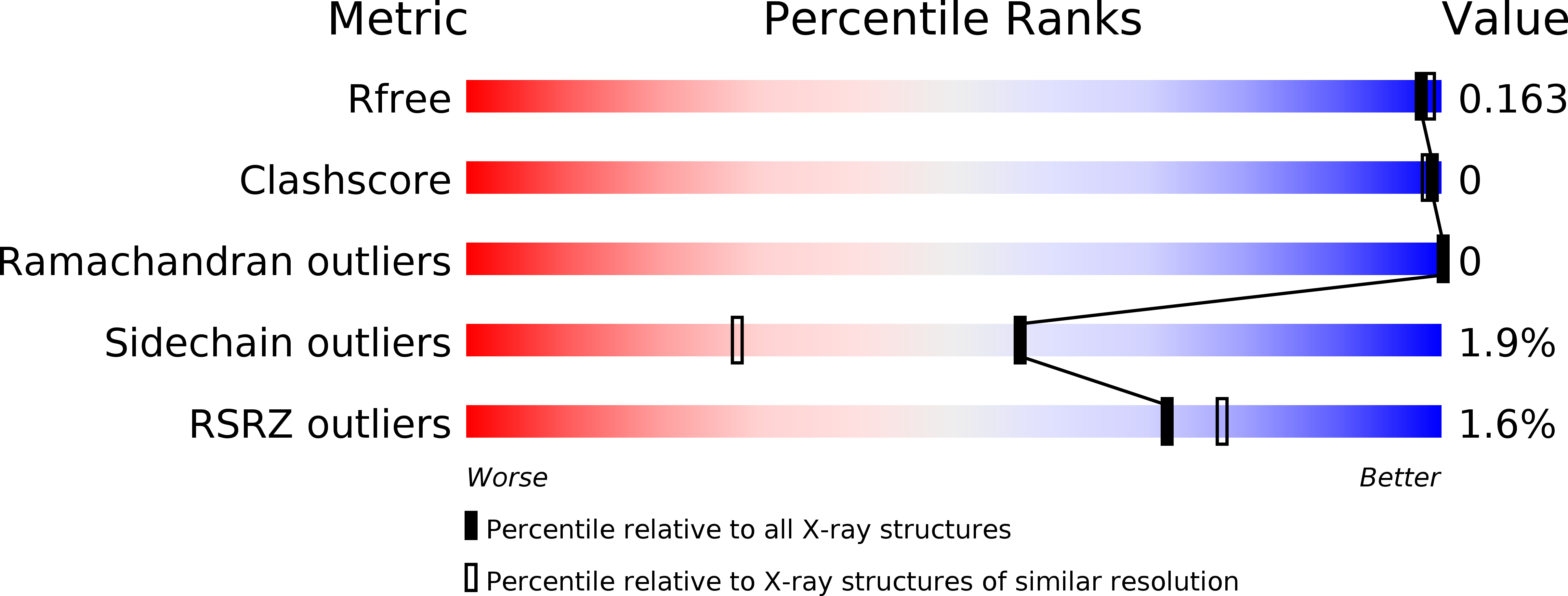
R-Value Free:
0.16
R-Value Work:
0.14
R-Value Observed:
0.14
Space Group:
P 31 2 1