
Deposition Date
2015-06-15
Release Date
2015-08-05
Last Version Date
2023-09-27
Entry Detail
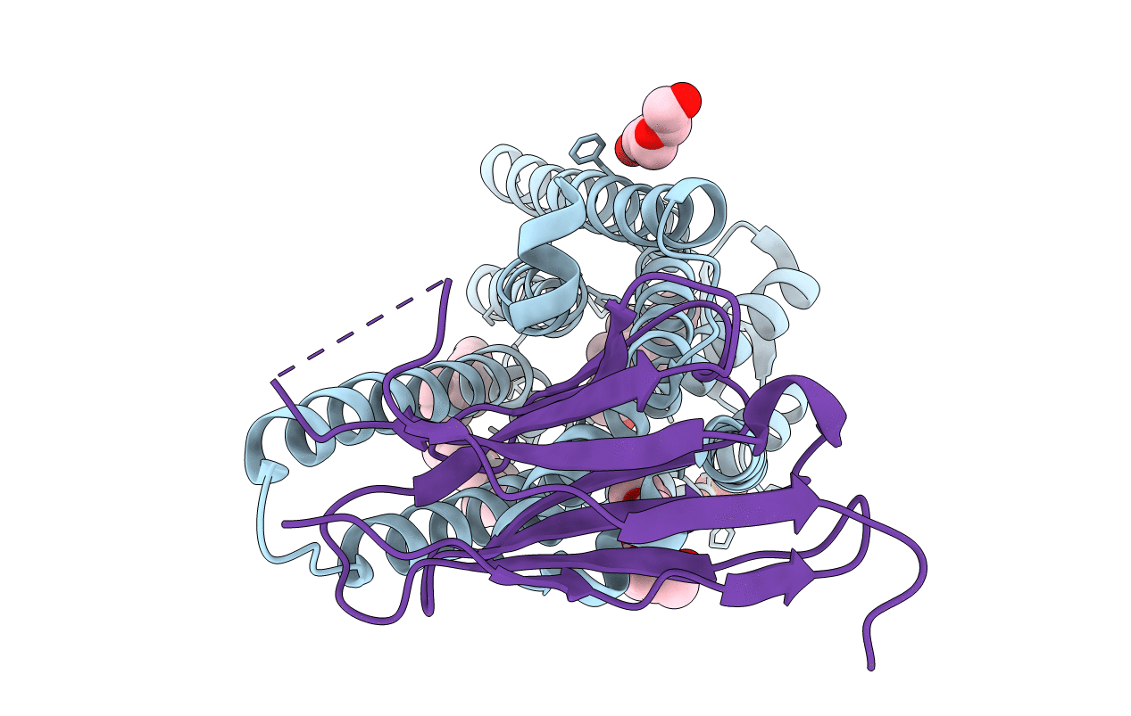
PDB ID:
5C1M
Keywords:
Title:
Crystal structure of active mu-opioid receptor bound to the agonist BU72
Biological Source:
Source Organism(s):
Mus musculus (Taxon ID: 10090)
Lama glama (Taxon ID: 9844)
Lama glama (Taxon ID: 9844)
Expression System(s):
Method Details:
Experimental Method:
Resolution:
2.07 Å
R-Value Free:
0.22
R-Value Work:
0.19
R-Value Observed:
0.20
Space Group:
I 21 21 21
