5C0Q image
Deposition Date 2015-06-12
Release Date 2015-09-16
Last Version Date 2024-10-30
Entry Detail
PDB ID:
5C0Q
Keywords:
Title:
Crystal structure of Zn bound CbsA from Thermotoga neapolitana
Biological Source:
Source Organism:
Host Organism:
Method Details:
Experimental Method:
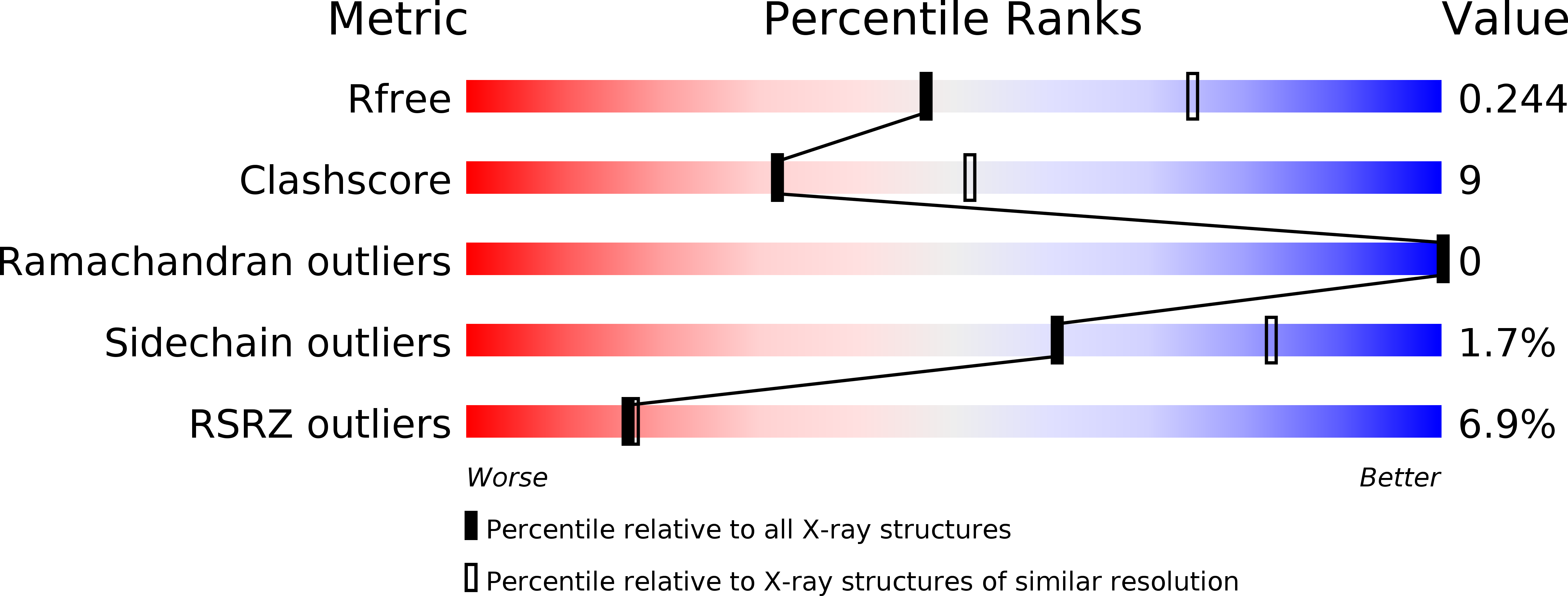
Resolution:
2.50 Å
R-Value Free:
0.24
R-Value Work:
0.20
R-Value Observed:
0.20
Space Group:
H 3 2
Macromolecular Entities
Polymer Type:polypeptide(L)
Molecule:Beta-N-acetylhexosaminidase
Gene (Uniprot):cbsA
Chain IDs:A, B, C, D
Chain Length:467
Number of Molecules:4
Biological Source:Thermotoga neapolitana
Ligand Molecules
Primary Citation
Crystal structure of beta-N-acetylglucosaminidase CbsA from Thermotoga neapolitana
Biochem.Biophys.Res.Commun. 464 869 874 (2015)
PMID: 26187666 DOI: 10.1016/j.bbrc.2015.07.053

Abstact

CbsA from the thermophilic marine bacteria Thermotoga neapolitana is a chitinolyitc enzyme that can cleave a glycosidic bond of the polymer N-acetylglucosamine at the non-reducing end. This enzyme has particularly high activity on di-N-acetylchitobiose. CbsA consists of a family of 3 glycoside hydrolase (GH3)-type catalytic domains and a unique C-terminal domain. The C-terminal domain distinguishes CbsA from other GH3-type enzymes. Sequence analyses have suggested that CbsA has the Asp-His dyad as a general acid/base with the NagZ of Bacillus subtilis and the Salmonella enterica serovar Typhimurium. Here, we determined the crystal structure of CbsA from T. neapolitana at a resolution of 2.0 Å using the Zn-SAD method, revealing a unique homodimeric assembly facilitated by the C-terminal domains in the dimer. We observed that CbsA is strongly inhibited by ZnCl2, and two zinc ions were consistently bound in the active site. Our results can explain the zinc ion's inhibition mechanism in the subfamily of GH3 enzymes, and provide information on the structural diversity and substrate specificity of this hydrolase family.

Legend

Protein

Chemical

Disease

Primary Citation of related structures