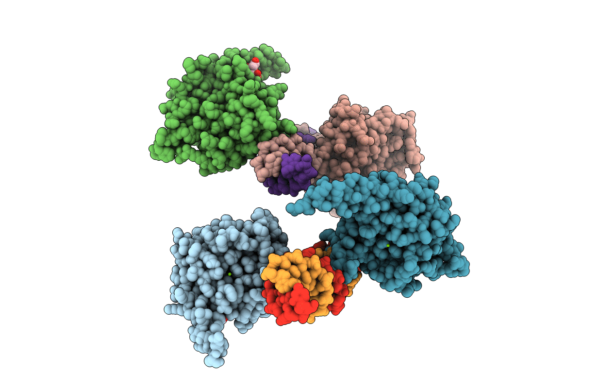
5BYG image
Deposition Date 2015-06-10
Release Date 2015-09-23
Last Version Date 2023-09-27
Entry Detail
PDB ID:
5BYG
Title:
X-ray structure of AAV2 OBD-AAVS1 complex 2:1
Biological Source:
Source Organism:
Host Organism:
Method Details:
Experimental Method:
Resolution:
2.50 Å
R-Value Free:
0.22
R-Value Work:
0.18
R-Value Observed:
0.19
Space Group:
C 1 2 1
Macromolecular Entities
Structures with similar UniProt ID
Protein Blast
Polymer Type:polypeptide(L)
Molecule:Protein Rep78
Gene (Uniprot):Rep78
Chain IDs:A, B, C, D
Chain Length:213
Number of Molecules:4
Biological Source:Adeno-associated virus 2
Polymer Type:polydeoxyribonucleotide
Molecule:DNA (5'-D(*CP*TP*CP*GP*GP*CP*GP*CP*TP*CP*GP*CP*TP*CP*GP*CP*TP*CP*GP*CP*T)-3')
Chain IDs:E, G
Chain Length:21
Number of Molecules:2
Biological Source:Homo sapiens
Polymer Type:polydeoxyribonucleotide
Molecule:DNA (5'-D(*GP*AP*GP*CP*GP*AP*GP*CP*GP*AP*GP*CP*GP*AP*GP*CP*GP*CP*CP*GP*A)-3')
Chain IDs:F, H
Chain Length:21
Number of Molecules:2
Biological Source:Homo sapiens
Primary Citation
Structural Insights into the Assembly of the Adeno-associated Virus Type 2 Rep68 Protein on the Integration Site AAVS1.
J.Biol.Chem. 290 27487 27499 (2015)
PMID: 26370092 DOI: 10.1074/jbc.M115.669960

Abstact

Adeno-associated virus (AAV) is the only eukaryotic virus with the property of establishing latency by integrating site-specifically into the human genome. The integration site known as AAVS1 is located in chromosome 19 and contains multiple GCTC repeats that are recognized by the AAV non-structural Rep proteins. These proteins are multifunctional, with an N-terminal origin-binding domain (OBD) and a helicase domain joined together by a short linker. As a first step to understand the process of site-specific integration, we proceeded to characterize the recognition and assembly of Rep68 onto the AAVS1 site. We first determined the x-ray structure of AAV-2 Rep68 OBD in complex with the AAVS1 DNA site. Specificity is achieved through the interaction of a glycine-rich loop that binds the major groove and an α-helix that interacts with a downstream minor groove on the same face of the DNA. Although the structure shows a complex with three OBD molecules bound to the AAVS1 site, we show by using analytical centrifugation and electron microscopy that the full-length Rep68 forms a heptameric complex. Moreover, we determined that a minimum of two direct repeats is required to form a stable complex and to melt DNA. Finally, we show that although the individual domains bind DNA poorly, complex assembly requires oligomerization and cooperation between its OBD, helicase, and the linker domains.

Legend

Protein

Chemical

Disease

Primary Citation of related structures