5BWZ image
Deposition Date 2015-06-08
Release Date 2016-06-08
Last Version Date 2023-09-27
Entry Detail
PDB ID:
5BWZ
Title:
Crystal structure of S39E HDAC8 in complex with Droxinostat
Biological Source:
Source Organism:
Homo sapiens (Taxon ID: 9606)
Host Organism:
Method Details:
Experimental Method:
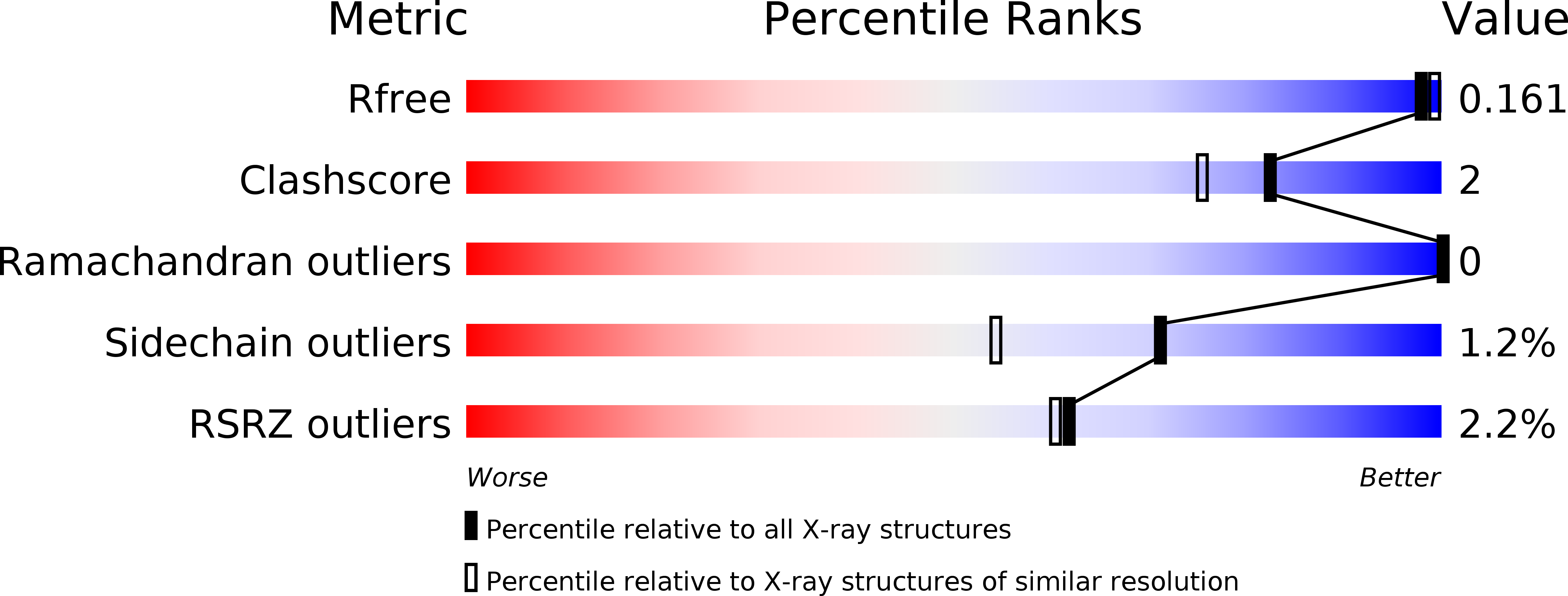
Resolution:
1.59 Å
R-Value Free:
0.16
R-Value Work:
0.14
R-Value Observed:
0.14
Space Group:
P 1 21 1
Macromolecular Entities
Structures with similar UniProt ID
Protein Blast
Polymer Type:polypeptide(L)
Molecule:Histone deacetylase 8
Gene (Uniprot):HDAC8
Mutagens:S39E
Chain IDs:A, B
Chain Length:389
Number of Molecules:2
Biological Source:Homo sapiens
Primary Citation
Phosphorylation of Histone Deacetylase 8: Structural and Mechanistic Analysis of the Phosphomimetic S39E Mutant.
Biochemistry 58 4480 4493 (2019)
PMID: 31633931 DOI: 10.1021/acs.biochem.9b00653

Abstact

Histone deacetylase (HDAC) enzymes that catalyze removal of acetyl-lysine post-translational modifications are frequently post-translationally modified. HDAC8 is phosphorylated within the deacetylase domain at conserved residue serine 39, which leads to decreased catalytic activity. HDAC8 phosphorylation at S39 is unique in its location and function and may represent a novel mode of deacetylation regulation. To better understand the impact of phosphorylation of HDAC8 on enzyme structure and function, we performed crystallographic, kinetic, and molecular dynamics studies of the S39E HDAC8 phosphomimetic mutant. This mutation decreases the level of deacetylation of peptides derived from acetylated nuclear and cytoplasmic proteins. However, the magnitude of the effect depends on the peptide sequence and the identity of the active site metal ion [Zn(II) vs Fe(II)], with the value of kcat/KM for the mutant decreasing 9- to >200-fold compared to that of wild-type HDAC8. Furthermore, the dissociation rate constant of the active site metal ion increases by ∼10-fold. S39E HDAC8 was crystallized in complex with the inhibitor Droxinostat, revealing that phosphorylation of S39, as mimicked by the glutamate side chain, perturbs local structure through distortion of the L1 loop. Molecular dynamics simulations of both S39E and phosphorylated S39 HDAC8 demonstrate that the perturbation of the L1 loop likely occurs because of the lost hydrogen bond between D29 and S39. Furthermore, the S39 perturbation causes structural changes that propagate through the protein scaffolding to influence function in the active site. These data demonstrate that phosphorylation plays an important regulatory role for HDAC8 by affecting ligand binding, catalytic efficiency, and substrate selectivity.

Legend

Protein

Chemical

Disease

Primary Citation of related structures