Deposition Date
2015-06-08
Release Date
2016-07-13
Last Version Date
2023-11-08
Entry Detail
PDB ID:
5BWN
Keywords:
Title:
Crystal Structure of SIRT3 with a H3K9 Peptide Containing a Myristoyl Lysine
Biological Source:
Source Organism(s):
Homo sapiens (Taxon ID: 9606)
synthetic construct (Taxon ID: 32630)
synthetic construct (Taxon ID: 32630)
Expression System(s):
Method Details:
Experimental Method:
Resolution:
1.94 Å
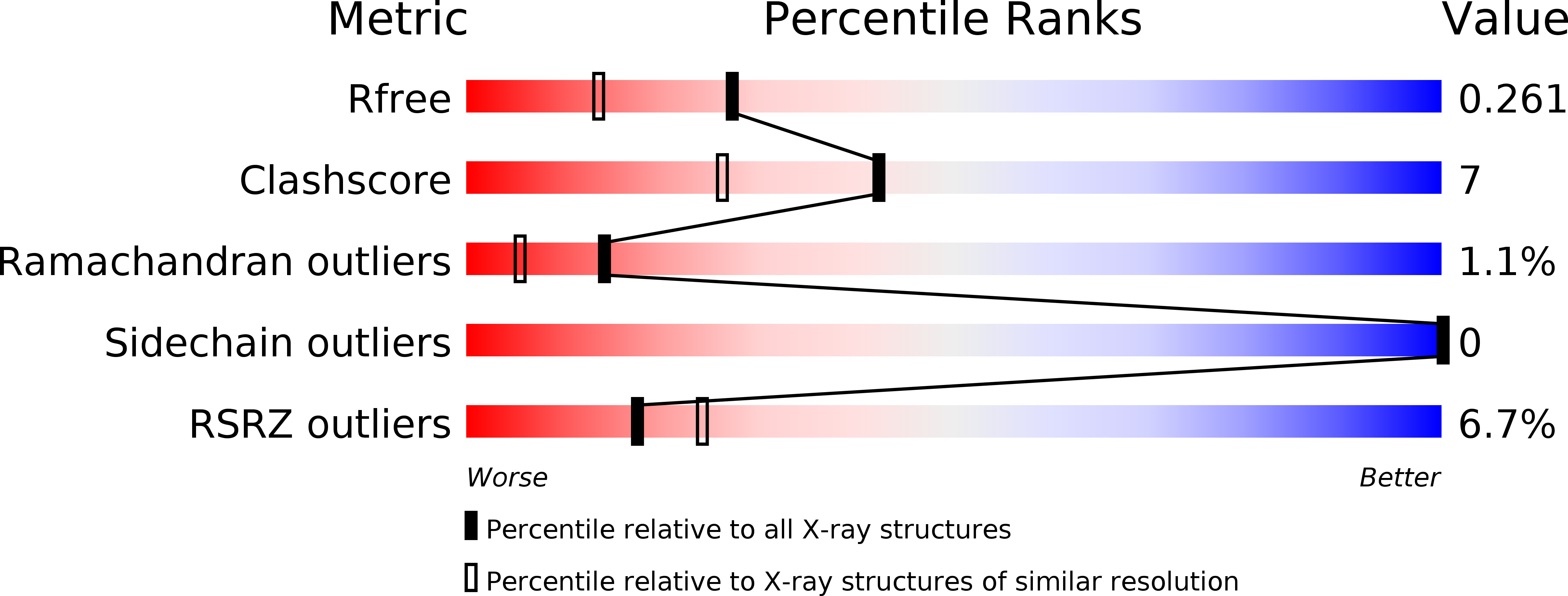
R-Value Free:
0.27
R-Value Work:
0.22
R-Value Observed:
0.23
Space Group:
P 21 21 21