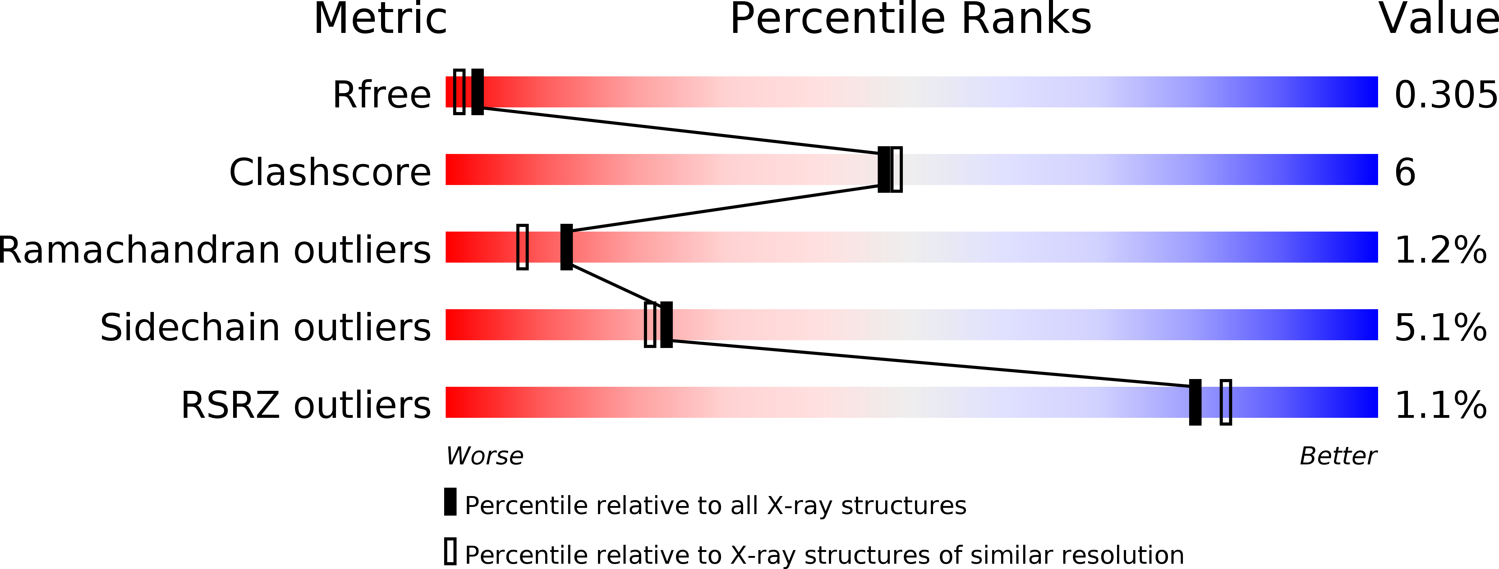
5BVQ image
Deposition Date 2015-06-05
Release Date 2015-08-05
Last Version Date 2024-03-20
Entry Detail
PDB ID:
5BVQ
Title:
Ligand-unbound pFABP4
Biological Source:
Source Organism:
Host Organism:
Method Details:
Experimental Method:
Resolution:
2.10 Å
R-Value Free:
0.29
R-Value Work:
0.24
R-Value Observed:
0.24
Space Group:
P 61
Macromolecular Entities
Polymer Type:polypeptide(L)
Molecule:fatty acid-binding protein
Chain IDs:A, B
Chain Length:132
Number of Molecules:2
Biological Source:Pygoscelis papua
Primary Citation
Structural basis for the ligand-binding specificity of fatty acid-binding proteins (pFABP4 and pFABP5) in gentoo penguin
Biochem.Biophys.Res.Commun. 465 12 18 (2015)
PMID: 26206084 DOI: 10.1016/j.bbrc.2015.07.087

Abstact

Fatty acid-binding proteins (FABPs) are involved in transporting hydrophobic fatty acids between various aqueous compartments of the cell by directly binding ligands inside their β-barrel cavities. Here, we report the crystal structures of ligand-unbound pFABP4, linoleate-bound pFABP4, and palmitate-bound pFABP5, obtained from gentoo penguin (Pygoscelis papua), at a resolution of 2.1 Å, 2.2 Å, and 2.3 Å, respectively. The pFABP4 and pFABP5 proteins have a canonical β-barrel structure with two short α-helices that form a cap region and fatty acid ligand binding sites in the hydrophobic cavity within the β-barrel structure. Linoleate-bound pFABP4 and palmitate-bound pFABP5 possess different ligand-binding modes and a unique ligand-binding pocket due to several sequence dissimilarities (A76/L78, T30/M32, underlining indicates pFABP4 residues) between the two proteins. Structural comparison revealed significantly different conformational changes in the β3-β4 loop region (residues 57-62) as well as the flipped Phe60 residue of pFABP5 than that in pFABP4 (the corresponding residue is Phe58). A ligand-binding study using fluorophore displacement assays shows that pFABP4 has a relatively strong affinity for linoleate as compared to pFABP5. In contrast, pFABP5 exhibits higher affinity for palmitate than that for pFABP4. In conclusion, our high-resolution structures and ligand-binding studies provide useful insights into the ligand-binding preferences of pFABPs based on key protein-ligand interactions.

Legend

Protein

Chemical

Disease

Primary Citation of related structures