
Deposition Date
2015-05-28
Release Date
2016-03-02
Last Version Date
2023-11-08
Entry Detail
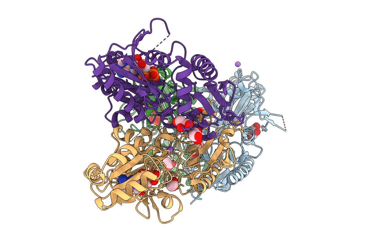
PDB ID:
5BPF
Keywords:
Title:
Crystal structure of ADP complexed D-alanine-D-alanine ligase(DDL) from Yersinia pestis
Biological Source:
Source Organism(s):
Yersinia pestis (Taxon ID: 632)
Expression System(s):
Method Details:
Experimental Method:
Resolution:
2.28 Å
R-Value Free:
0.25
R-Value Work:
0.18
R-Value Observed:
0.18
Space Group:
P 21 21 21
