
Deposition Date
2015-05-24
Release Date
2015-07-29
Last Version Date
2023-11-08
Entry Detail
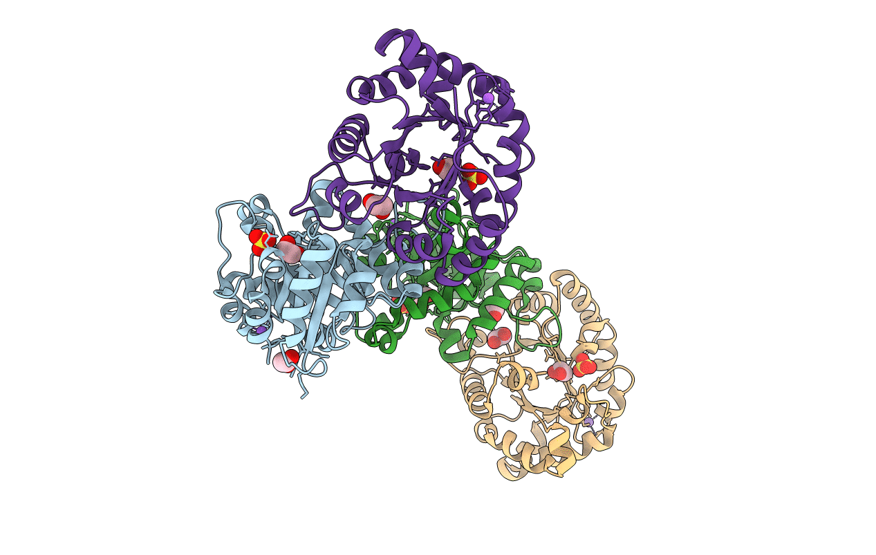
PDB ID:
5BMX
Keywords:
Title:
Crystal structure of T75N mutant of Triosephosphate isomerase from Plasmodium falciparum
Biological Source:
Source Organism(s):
Plasmodium falciparum (Taxon ID: 5833)
Expression System(s):
Method Details:
Experimental Method:
Resolution:
1.80 Å
R-Value Free:
0.21
R-Value Work:
0.16
R-Value Observed:
0.16
Space Group:
P 21 21 21
