Deposition Date
2016-06-14
Release Date
2016-10-26
Last Version Date
2024-11-20
Entry Detail
PDB ID:
5B8C
Keywords:
Title:
High resolution structure of the human PD-1 in complex with pembrolizumab Fv
Biological Source:
Source Organism(s):
Homo sapiens (Taxon ID: 9606)
Expression System(s):
Method Details:
Experimental Method:
Resolution:
2.15 Å
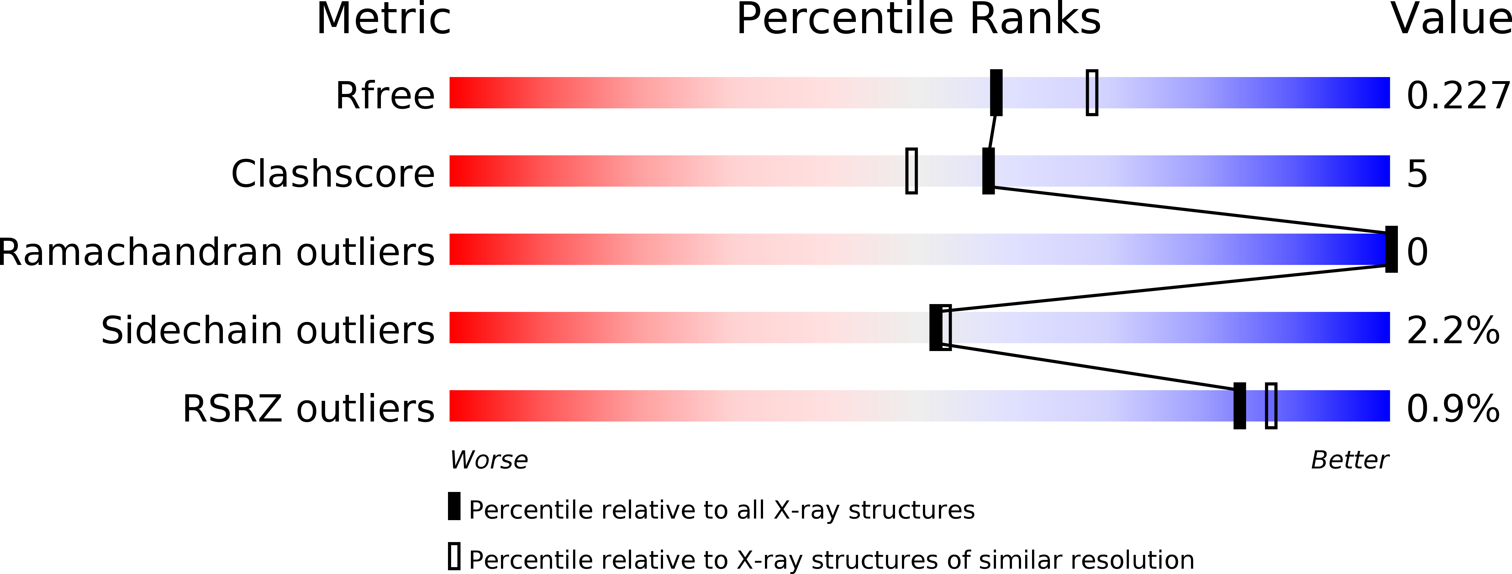
R-Value Free:
0.22
R-Value Work:
0.18
R-Value Observed:
0.18
Space Group:
P 21 21 21