
Deposition Date
2016-05-31
Release Date
2016-09-07
Last Version Date
2023-11-08
Entry Detail
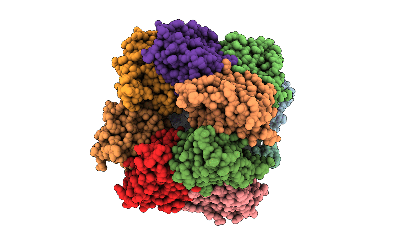
PDB ID:
5B6P
Keywords:
Title:
Structure of the dodecameric type-II dehydrogenate dehydratase from Acinetobacter baumannii at 2.00 A resolution
Biological Source:
Source Organism(s):
Acinetobacter baumannii (Taxon ID: 400667)
Expression System(s):
Method Details:
Experimental Method:
Resolution:
2.00 Å
R-Value Free:
0.23
R-Value Work:
0.20
R-Value Observed:
0.20
Space Group:
P 1 21 1
