5B6C image
Deposition Date 2016-05-26
Release Date 2017-01-04
Last Version Date 2024-03-20
Entry Detail
PDB ID:
5B6C
Title:
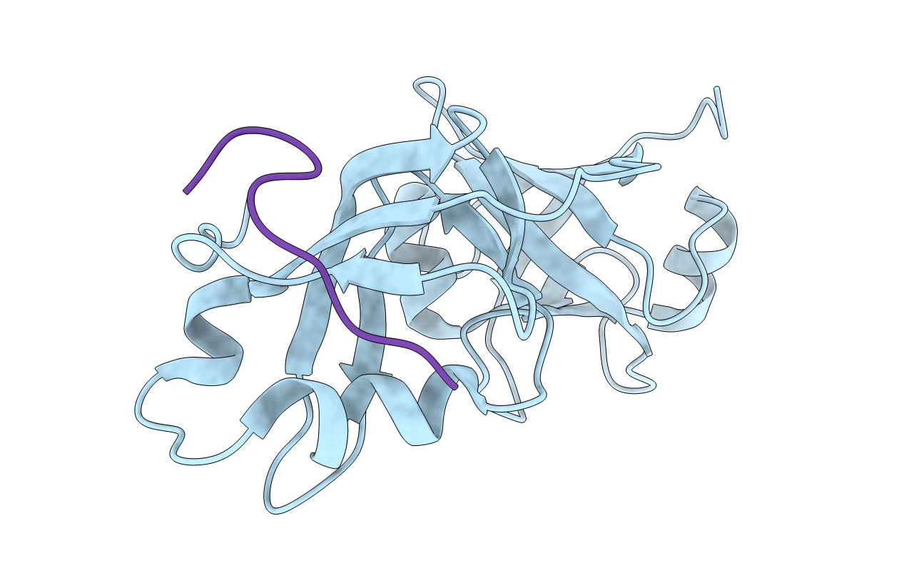
Structural Details of Ufd1 binding to p97
Biological Source:
Source Organism:
Homo sapiens (Taxon ID: 9606)
Host Organism:
Method Details:
Experimental Method:
Resolution:
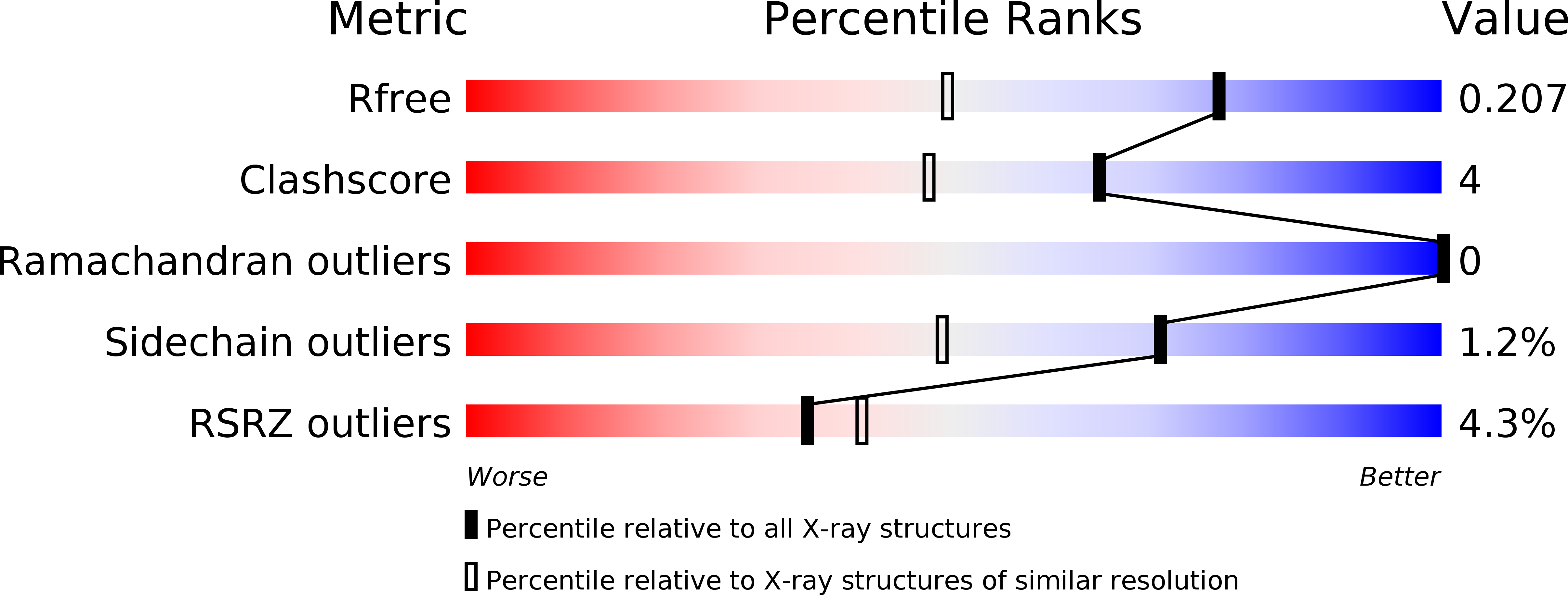
1.55 Å
R-Value Free:
0.19
R-Value Work:
0.15
R-Value Observed:
0.16
Space Group:
H 3
Macromolecular Entities
Structures with similar UniProt ID
Protein Blast
Polymer Type:polypeptide(L)
Molecule:Transitional endoplasmic reticulum ATPase
Gene (Uniprot):VCP
Chain IDs:A
Chain Length:174
Number of Molecules:1
Biological Source:Homo sapiens
Structures with similar UniProt ID
Protein Blast
Polymer Type:polypeptide(L)
Molecule:Peptide from Ubiquitin fusion degradation protein 1 homolog
Chain IDs:B
Chain Length:11
Number of Molecules:1
Biological Source:Homo sapiens
Primary Citation
Structural Details of Ufd1 Binding to p97 and Their Functional Implications in ER-Associated Degradation
PLoS ONE 11 e0163394 e0163394 (2016)
PMID: 27684549 DOI: 10.1371/journal.pone.0163394

Abstact

The hexameric ATPase p97 has been implicated in diverse cellular processes through interactions with many different adaptor proteins at its N-terminal domain. Among these, the Ufd1-Npl4 heterodimer is a major adaptor, and the p97-Ufd1-Npl4 complex plays an essential role in endoplasmic reticulum-associated degradation (ERAD), acting as a segregase that translocates the ubiquitinated client protein from the ER membrane into the cytosol for proteasomal degradation. We determined the crystal structure of the complex of the N-terminal domain of p97 and the SHP box of Ufd1 at a resolution of 1.55 Å. The 11-residue-long SHP box of Ufd1 binds at the far-most side of the Nc lobe of the p97 N domain primarily through hydrophobic interactions, such that F225, F228, N233 and L235 of the SHP box contact hydrophobic residues on the surface of the p97 Nc lobe. Mutating these key interface residues abolished the interactions in two different binding experiments, isothermal titration calorimetry and co-immunoprecipitation. Furthermore, cycloheximide chase assays showed that these same mutations caused accumulation of tyrosinase-C89R, a well-known ERAD substrate, thus implying decreased rate of protein degradation due to their defects in ERAD function. Together, these results provide structural and biochemical insights into the interaction between p97 N domain and Ufd1 SHP box.

Legend

Protein

Chemical

Disease

Primary Citation of related structures