5B4N image
Deposition Date 2016-04-06
Release Date 2016-09-14
Last Version Date 2023-11-08
Entry Detail
PDB ID:
5B4N
Keywords:
Title:
Structure analysis of function associated loop mutant of substrate recognition domain of Fbs1 ubiquitin ligase
Biological Source:
Source Organism:
Mus musculus (Taxon ID: 10090)
Method Details:
Experimental Method:
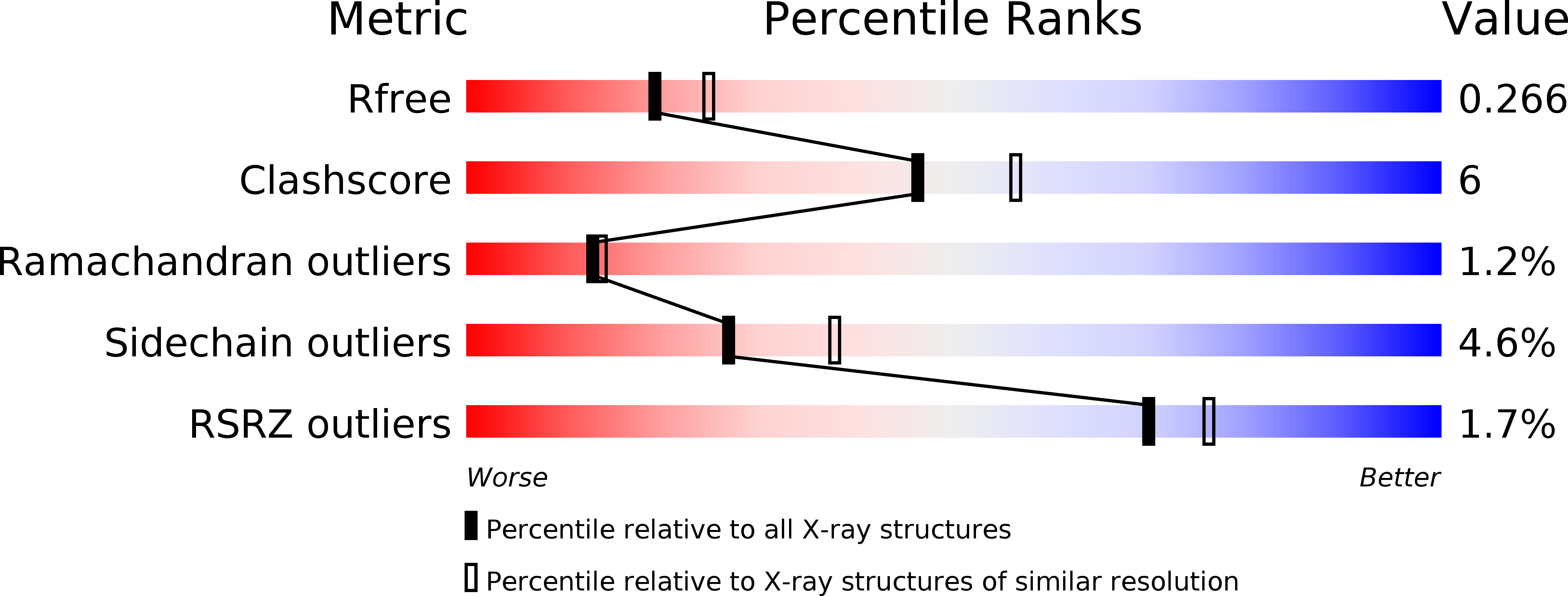
Resolution:
2.30 Å
R-Value Free:
0.26
R-Value Work:
0.18
R-Value Observed:
0.18
Space Group:
P 1 21 1
Macromolecular Entities
Protein Blast
Polymer Type:polypeptide(L)
Molecule:F-box only protein 2
Gene (Uniprot):Fbxo2
Chain IDs:A, B
Chain Length:203
Number of Molecules:2
Biological Source:Mus musculus
Primary Citation
Structural analysis of a function-associated loop mutant of the substrate-recognition domain of Fbs1 ubiquitin ligase
Acta Crystallogr.,Sect.F 72 619 626 (2016)
PMID: 27487926 DOI: 10.1107/S2053230X16011018

Abstact

The SCF ubiquitin ligase comprises four components: Skp1, Cul1, Rbx1 and a variable-subunit F-box protein. The F-box protein Fbs1, which recognizes the N-linked glycoproteins, is involved in the endoplasmic reticulum-associated degradation pathway. Although FBG3, another F-box protein, shares 51% sequence identity with Fbs1, FBG3 does not bind glycoproteins. To investigate the sequence-structure relationship of the substrate-binding pocket, the crystal structure of a mutant substrate-binding domain of Fbs1 in which the six nonconserved regions (β1, β2-β3, β3-β4, β5-β6, β7-β8 and β9-β10) of Fbs1 were substituted with those of FBG3 was determined. The substrate-binding pocket of this model exhibits structural features that differ from those of Fsb1.

Legend

Protein

Chemical

Disease

Primary Citation of related structures
Feedback Form
Name
Email
Institute
Feedback