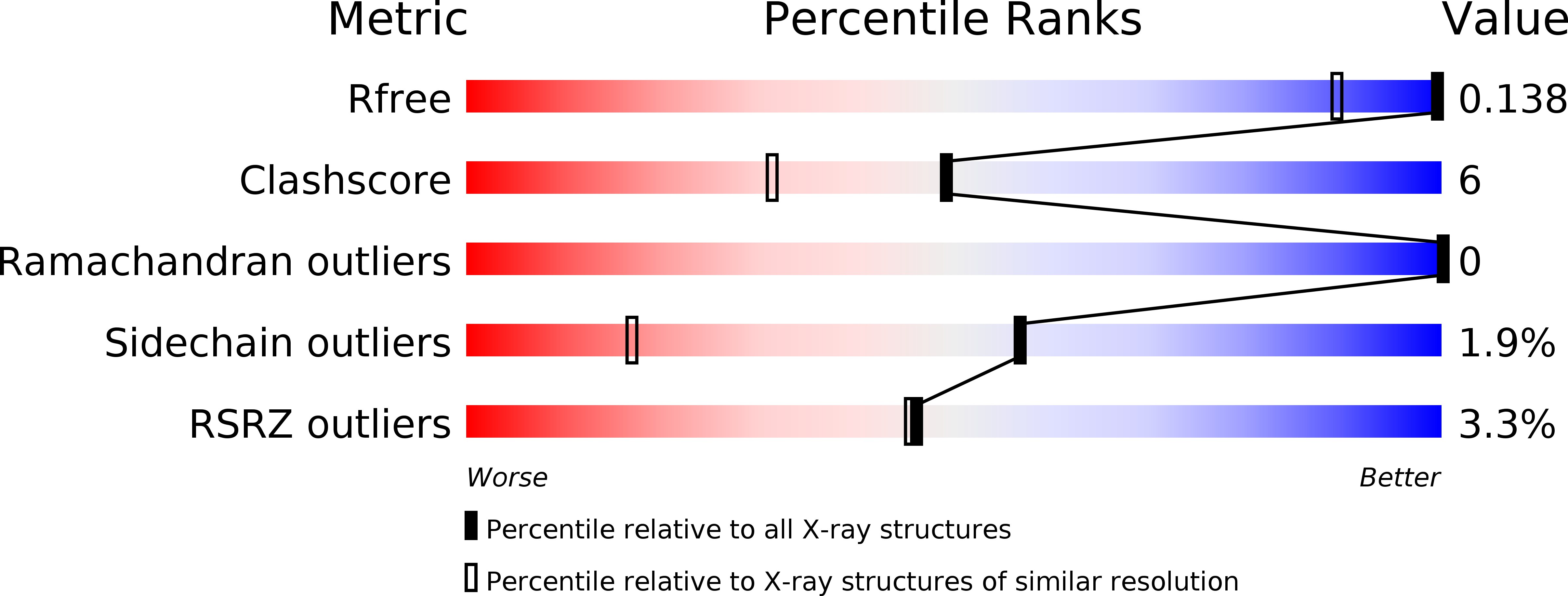
5B4I image
Deposition Date 2016-04-04
Release Date 2017-03-15
Last Version Date 2023-11-08
Entry Detail
PDB ID:
5B4I
Keywords:
Title:
Crystal structure of I86D mutant of phycocyanobilin:ferredoxin oxidoreductase in complex with biliverdin (data 2)
Biological Source:
Source Organism:
Method Details:
Experimental Method:
Resolution:
1.11 Å
R-Value Free:
0.15
R-Value Work:
0.12
Space Group:
P 21 21 2
Macromolecular Entities
Polymer Type:polypeptide(L)
Molecule:Phycocyanobilin:ferredoxin oxidoreductase
Gene (Uniprot):pcyA
Mutations:I86D
Chain IDs:A
Chain Length:248
Number of Molecules:1
Biological Source:Synechocystis sp. PCC 6803
Ligand Molecules
Primary Citation
Atomic-resolution structure of the phycocyanobilin:ferredoxin oxidoreductase I86D mutant in complex with fully protonated biliverdin
FEBS Lett. 590 3425 3434 (2016)
PMID: 27596987 DOI: 10.1002/1873-3468.12387

Abstact

Phycocyanobilin:ferredoxin oxidoreductase (PcyA) catalyzes the reduction of biliverdin (BV) to produce phycocyanobilin, a linear tetrapyrrole pigment used for light harvesting and light sensing. Spectroscopic and HPLC analyses inidicate that BV bound to the I86D mutant of PcyA is fully protonated (BVH+) and can accept an electron, but I86D is unable to donate protons for the reduction; therefore, compared to the wild-type PcyA, the I86D mutant stabilizes BVH+ . To elucidate the structural basis of the I86D mutation, we determined the atomic-resolution structure of the I86D-BVH+ complex and the protonation states of the essential residues Asp105 and Glu76 in PcyA. Our study revealed that Asp105 adopted a fixed conformation in the I86D mutant, although it had dual conformations in wild-type PcyA which reflected the protonation states of BV. Taken together with biochemical/spectroscopic results, our analysis of the I86D-BVH+ structure supports the hypothesis that flexibility of Asp105 is essential for the catalytic activity of PcyA.

Legend

Protein

Chemical

Disease

Primary Citation of related structures