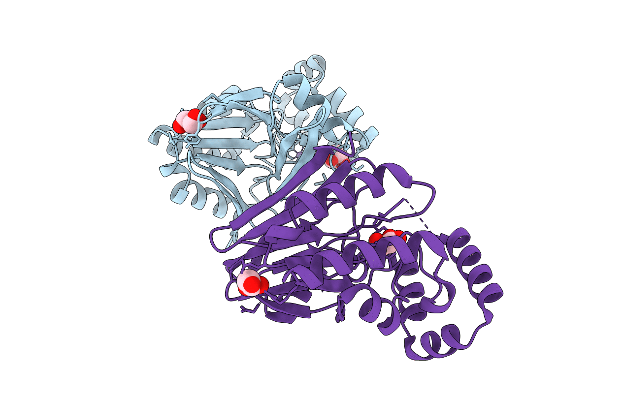
5B4C image
Entry Detail
PDB ID:
5B4C
Keywords:
Title:
Crystal structure of H10N mutant of LpxH with manganese
Biological Source:
Host Organism:
PDB Version:
Deposition Date:
2016-04-03
Release Date:
2016-09-28
Method Details:
Experimental Method:
Resolution:
1.96 Å
R-Value Free:
0.21
R-Value Work:
0.17
R-Value Observed:
0.17
Space Group:
P 21 21 21
Macromolecular Entities
Polymer Type:polypeptide(L)
Description:UDP-2,3-diacylglucosamine hydrolase
Mutations:H10N
Chain IDs:A, B
Chain Length:248
Number of Molecules:2
Biological Source:Pseudomonas aeruginosa PAO1
Primary Citation
Crystal structures of the UDP-diacylglucosamine pyrophosphohydrase LpxH from Pseudomonas aeruginosa
Sci Rep 6 32822 32822 (2016)
PMID: 27609419 DOI: 10.1038/srep32822

Abstact

Lipid A (also known as endotoxin) is the hydrophobic portion of lipopolysaccharides. It is an essential membrane component required for the viability of gram-negative bacteria. The enzymes involved in its biosynthesis are attractive targets for the development of novel antibiotics. LpxH catalyzes the fourth step of the lipid A biosynthesis pathway and cleaves the pyrophosphate bond of UDP-2,3-diacylglucosamine to yield 2,3-diacylglucosamine 1-phosphate (lipid X) and UMP. Here we present the structures of LpxH from Pseudomonas aeruginosa (PaLpxH). PaLpxH consists of two domains: a catalytic domain that is homologous to the metallophosphoesterases and a helical insertion domain. Lipid X was captured in the crevice between these two domains, with its phosphate group facing the dinuclear metal (Mn(2+)) center and two acyl chains buried in the hydrophobic cavity. The structures reveal that a large conformational change occurs at the lipid X binding site surface upon the binding/release of the product molecule. Based on these observations, we propose a novel model for lipid X embedding, which involves the scissor-like movement of helix α6, resulting in the release of lipid X into the lipid bilayer.

Legend

Protein

Chemical

Disease

Primary Citation of related structures