5B47 image
Entry Detail
PDB ID:
5B47
Keywords:
Title:
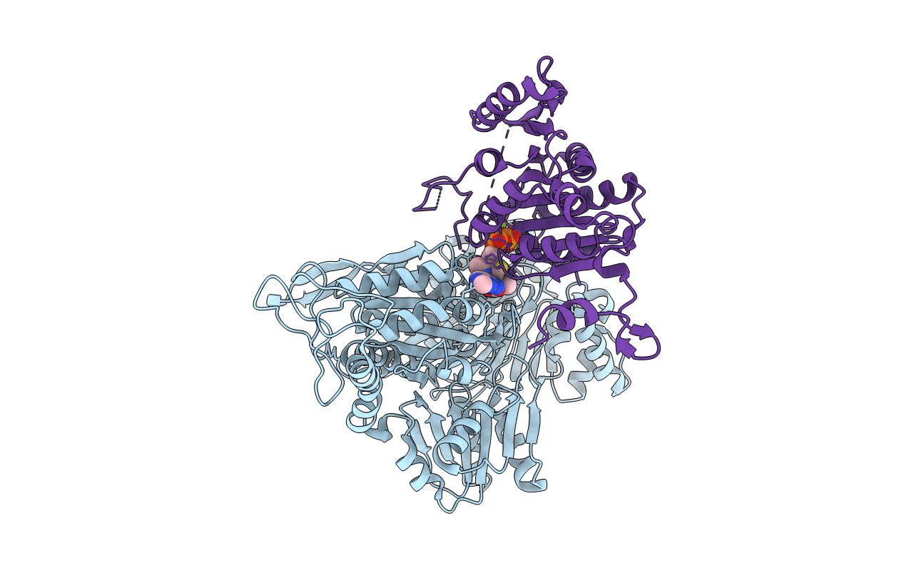
2-Oxoacid:Ferredoxin Oxidoreductase 2 from Sulfolobus tokodai - pyruvate complex
Biological Source:
Host Organism:
PDB Version:
Deposition Date:
2016-04-01
Release Date:
2016-09-28
Method Details:
Experimental Method:
Resolution:
2.20 Å
R-Value Free:
0.25
R-Value Work:
0.20
R-Value Observed:
0.20
Space Group:
C 2 2 21
Macromolecular Entities
Polymer Type:polypeptide(L)
Description:2-oxoacid--ferredoxin oxidoreductase alpha subunit
Chain IDs:A
Chain Length:628
Number of Molecules:1
Biological Source:Sulfolobus tokodaii str. 7
Polymer Type:polypeptide(L)
Description:2-oxoacid--ferredoxin oxidoreductase beta subunit
Chain IDs:B
Chain Length:304
Number of Molecules:1
Biological Source:Sulfolobus tokodaii str. 7
Primary Citation
Crystal structures of archaeal 2-oxoacid:ferredoxin oxidoreductases from Sulfolobus tokodaii
Sci Rep 6 33061 33061 (2016)
PMID: 27619895 DOI: 10.1038/srep33061

Abstact

As the first three-dimensional structure of the two-subunit type 2-oxoacid:ferredoxin oxidoreductases (OFOR) from archaea, we solved the crystal structures of STK_23000/STK_22980 (StOFOR1) and STK_24350/STK_24330 (StOFOR2) from Sulfolobus tokodaii. They showed similar overall structures, consisting of two a- and b-subunit heterodimers containing thiamin pyrophosphate (TPP) cofactor and [4Fe-4S] cluster, but lack an intramolecular ferredoxin domain. Unlike other OFORs, StOFORs can utilize both pyruvate and 2-oxoglutarate, playing a key role in the central metabolism. In the structure of StOFOR2 in unreacted pyruvate complex form, carboxylate group of pyruvate is recognized by Arg344 and Thr257 from the a-subunit, which are conserved in pyruvate:ferredoxin oxidoreductase from Desulfovbrio africanus (DaPFOR). In the structure of StOFOR1 co-crystallized with 2-oxobutyrate, electron density corresponding to a 1-hydroxypropyl group (post-decarboxylation state) was observed at the thiazole ring of TPP. The binding pockets of the StOFORs surrounding the methyl or propyl group of the ligands are wider than that of DaPFOR. Mutational analyses indicated that several residues were responsible for the broad 2-oxoacid specificity of StOFORs. We also constructed a possible complex structural model by placing a Zn(2+)-containing dicluster ferredoxin of S. tokodaii into the large pocket of StOFOR2, providing insight into the electron transfer between the two redox proteins.

Legend

Protein

Chemical

Disease

Primary Citation of related structures