5B3V image
Deposition Date 2016-03-13
Release Date 2017-02-15
Last Version Date 2023-11-08
Entry Detail
PDB ID:
5B3V
Keywords:
Title:
Crystal structure of biliverdin reductase in complex with biliverdin and NADP+ from Synechocystis sp. PCC 6803
Biological Source:
Source Organism:
Host Organism:
Method Details:
Experimental Method:
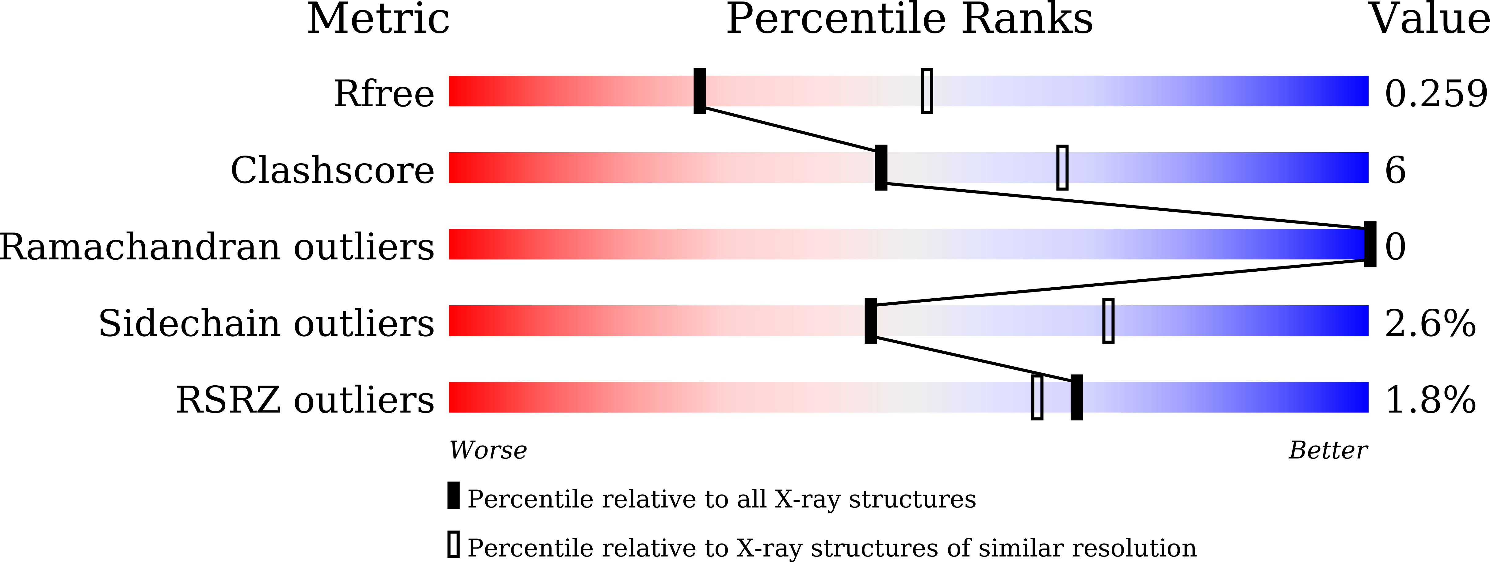
Resolution:
2.59 Å
R-Value Free:
0.25
R-Value Work:
0.20
R-Value Observed:
0.21
Space Group:
P 1 21 1
Macromolecular Entities
Polymer Type:polypeptide(L)
Molecule:Biliverdin reductase
Gene (Uniprot):bvdR
Chain IDs:A (auth: B), B (auth: A), C, D
Chain Length:331
Number of Molecules:4
Biological Source:Synechocystis sp.
Primary Citation
A substrate-bound structure of cyanobacterial biliverdin reductase identifies stacked substrates as critical for activity
Nat Commun 8 14397 14397 (2017)
PMID: 28169272 DOI: 10.1038/ncomms14397

Abstact

Biliverdin reductase catalyses the last step in haem degradation and produces the major lipophilic antioxidant bilirubin via reduction of biliverdin, using NAD(P)H as a cofactor. Despite the importance of biliverdin reductase in maintaining the redox balance, the molecular details of the reaction it catalyses remain unknown. Here we present the crystal structure of biliverdin reductase in complex with biliverdin and NADP+. Unexpectedly, two biliverdin molecules, which we designated the proximal and distal biliverdins, bind with stacked geometry in the active site. The nicotinamide ring of the NADP+ is located close to the reaction site on the proximal biliverdin, supporting that the hydride directly attacks this position of the proximal biliverdin. The results of mutagenesis studies suggest that a conserved Arg185 is essential for the catalysis. The distal biliverdin probably acts as a conduit to deliver the proton from Arg185 to the proximal biliverdin, thus yielding bilirubin.

Legend

Protein

Chemical

Disease

Primary Citation of related structures